
Cardiology Made Simple® Clinical Handbook Series · Volume I
Online Reading Edition · First online release: 3 November 2026
This edition is available for online reading only. Downloading and printing are not enabled.
Developed in accordance with the UVP INTELLECTUS® Editorial Excellence Standard.
Abstract
A physiology-based approach to ECG interpretation and clinical reasoning.
This handbook explains how electrical activation normally travels through the heart and how disturbances in that activation produce a wide QRS complex. It begins with the clinical importance of recognising wide-QRS tachyarrhythmias, then follows the normal conduction pathway from the sinoatrial node through the atrioventricular node to the bundle of His, the bundle branches and the Purkinje network, and explains why the normal QRS complex is narrow. It then describes five principal mechanisms of QRS widening: bundle branch or fascicular conduction abnormality, ventricular-origin activation, ventricular pre-excitation, ventricular pacing and diffuse intraventricular conduction slowing. Interpretation is organised around four questions: where did the impulse originate, which pathway did it follow, how rapidly did it propagate and how did the activation sequence change? Throughout, the volume emphasises that a wide QRS is an observation rather than a diagnosis, that haemodynamic stability does not exclude ventricular tachycardia, and that immediate patient safety takes priority over diagnostic certainty. It is an evidence-informed educational resource based on a narrative synthesis of the literature reviewed to September 2026; it is not a systematic review or a clinical practice guideline.
Keywords: Cardiac conduction system; His–Purkinje system; QRS complex; Wide-QRS tachycardia; Ventricular activation; ECG interpretation; Clinical reasoning; Cardiology education.
Publication Information
| Series | Cardiology Made Simple® Clinical Handbook Series |
|---|---|
| Volume I | The Electrophysiological Foundations |
| Subtitle | From Normal Cardiac Conduction to Wide-QRS Tachyarrhythmias |
| Author | Mark C. Amadi |
| External Clinical Reviewer | Dr Ebenezer Wireko, MBChB, MPH, MPhil, MAM Clinical Director, Mamprobi Hospital |
| Publisher | Unveiling Voice Publications® (UVP) |
| Edition | First Edition, 2026 |
| First online release | 3 November 2026 |
| Internal publication reference | UVP-CMS-2026-V01 |
| ISSN | Application pending |
| Publisher’s website | www.unvo-ac.uk |
| cardiology@unvo-ac.uk |
This volume forms part of the Cardiology Made Simple® Clinical Handbook Series, published by Unveiling Voice Publications®. The series ISSN and associated bibliographic metadata will be updated following confirmation by the relevant registration authority. The internal publication reference is a publisher-assigned identifier and does not constitute a registered Digital Object Identifier (DOI).
Copyright
Copyright © 2026 Mark C. Amadi. All rights reserved.
No part of this publication may be reproduced, stored, transmitted or distributed in any form or by any means without prior written permission from the copyright holder, except as permitted under applicable copyright law.
Every effort has been made to ensure that this handbook reflects the scientific evidence and professional guidance available at the time of publication. However, medical knowledge, clinical recommendations and therapeutic practices continue to evolve.
This handbook is intended for education and professional development. It does not replace individual clinical judgement, current clinical guidelines, prescribing information, institutional policies or appropriate specialist consultation. Readers should consult current authoritative guidance and relevant clinical protocols when making decisions concerning patient care.
The author and publisher accept no responsibility for loss, injury or damage arising from the use or misuse of this publication, to the extent permitted by applicable law.
Dedication
To every healthcare professional whose commitment to learning improves the lives of others.
May this handbook strengthen your clinical reasoning, deepen your understanding, and inspire excellence in patient-centred cardiovascular care.
About the Author
Mark C. Amadi, PhD, MPH, MSc, is a public health researcher, cardiovascular health scholar, author and healthcare chaplain. His interdisciplinary interests encompass preventive cardiology, cardiovascular epidemiology, public health and clinical education.
He is the originator of the Triple-Risk Convergence Framework (TRCF) and producer of Cardiology Made Simple®. His publishing work is developed through UVP INTELLECTUS® | Unveiling Voice Publications®, with an emphasis on clinically grounded education and knowledge translation.
ORCID: 0009-0006-9502-9552 · Web of Science ResearcherID: GK-4948-2026 · Scopus Author ID: 60916734300 · SSRN Author ID: 13593028
Preface
Why This Handbook Was Written
Understanding cardiac arrhythmias begins with understanding how the normal heart generates and conducts electrical impulses. The electrocardiogram provides essential information about cardiac electrical activity, but meaningful interpretation requires an appreciation of the physiological mechanisms responsible for its waveforms and intervals.
This handbook was developed to strengthen that foundation.
Volume I of Cardiology Made Simple® examines normal cardiac impulse generation, atrioventricular conduction, the His–Purkinje system and the mechanisms governing ventricular activation. It then considers how disturbances in the origin, pathway, speed or sequence of electrical activation may alter the QRS complex and contribute to wide-QRS tachyarrhythmias.
The emphasis is on developing physiological understanding and structured clinical reasoning. Rather than encouraging reliance on ECG pattern recognition alone, the handbook invites readers to consider why an electrical abnormality occurs and what its underlying mechanism may mean for subsequent clinical assessment.
The wider Cardiology Made Simple® series will build upon these foundations through more detailed examination of diagnostic differentiation, clinical management and cardiovascular prevention.
The educational principle remains straightforward: make cardiovascular science easier to understand without compromising scientific accuracy.
Mark C. Amadi
How to Use This Handbook
A Guide to the CMS Educational Framework
Cardiology Made Simple® follows a progressive educational approach that connects normal physiology with abnormal electrical mechanisms and clinical reasoning.
Volume I begins with the normal cardiac conduction system before examining ventricular activation, QRS formation and the physiological mechanisms responsible for QRS widening.
Figures provide visual explanations of relevant anatomical and electrophysiological concepts. CMS Clinical Pearls® reinforce selected learning points, while chapter summaries and review materials support consolidation.
Four recurring questions guide interpretation:
Where did the impulse originate? → Which pathway did it follow? → How rapidly did it propagate? → How did the activation sequence change?
These questions encourage readers to move beyond describing ECG appearances towards understanding the mechanisms that produce them.
The CMS Learning Sequence™
UNDERSTAND → RECOGNISE → REASON → DIFFERENTIATE → ACT → PREVENT
This sequence represents the broader educational approach of the Cardiology Made Simple® series. Volume I concentrates on physiological understanding, recognition of altered ventricular activation and the foundations of clinical reasoning. Subsequent volumes will develop diagnostic differentiation, intervention and longer-term cardiovascular management in greater detail.
CMS Educational Principle®
Understand the physiology. Recognise the pattern. Reason clinically. Apply the evidence. Protect the patient.
Evidence and Editorial Methodology
Scientific Foundation of Cardiology Made Simple®
Cardiology Made Simple® is an evidence-informed educational handbook developed through a narrative synthesis of relevant cardiovascular literature, including professional clinical guidelines, scientific statements, reviews and peer-reviewed publications.
Priority has been given to authoritative guidance and established electrophysiological principles, with the evidence base reviewed to September 2026. The manuscript does not claim to constitute a systematic review or formal clinical practice guideline.
Vancouver referencing is used, with citation numbering restarting within each chapter and a corresponding reference list provided at the end of each chapter.
Figures and educational frameworks are intended to support conceptual understanding and clinical reasoning. Their inclusion should not be interpreted as evidence of independently validated educational effectiveness.
Generative artificial intelligence assisted selected aspects of editorial and educational development but was not used as an authoritative clinical source. The author retains responsibility for verifying scientific claims, references and the accuracy of the final manuscript. Further information is provided in the Generative Artificial Intelligence Transparency Statement.
The author retains responsibility for the scientific accuracy, editorial quality and final approval of this edition.
CMS Evidence Principle®
Simplify the science, never the standard.
Abbreviations
The following abbreviations are used throughout this handbook. Readers are encouraged to become familiar with these terms before progressing through the chapters.
| Abbreviation | Definition |
|---|---|
| ACC | American College of Cardiology |
| AF | Atrial fibrillation |
| AHA | American Heart Association |
| AV | Atrioventricular |
| AVNRT | Atrioventricular nodal re-entrant tachycardia |
| AVRT | Atrioventricular re-entrant tachycardia |
| BBB | Bundle branch block |
| bpm | Beats per minute |
| CMS | Cardiology Made Simple® |
| ECG | Electrocardiogram |
| ESC | European Society of Cardiology |
| ICD | Implantable cardioverter-defibrillator |
| IV | Intravenous |
| LBBB | Left bundle branch block |
| LV | Left ventricle; left ventricular |
| PR | PR interval |
| PVC | Premature ventricular complex |
| QRS | QRS complex |
| RBBB | Right bundle branch block |
| SA | Sinoatrial |
| SCD | Sudden cardiac death |
| SVT | Supraventricular tachycardia |
| UVP | Unveiling Voice Publications® |
| VT | Ventricular tachycardia |
| WPW | Wolff–Parkinson–White syndrome |
List of Figures
- Figure 1.1 The CMS Clinical Continuum™: From Recognition to Definitive Management
- Figure 2.1 The Normal Cardiac Electrical Conduction System
- Figure 2.2 The Sinoatrial Node: The Heart's Natural Pacemaker
- Figure 2.3 The Atrioventricular Node: The Heart's Physiological Gatekeeper
- Figure 3.1 The Bundle of His: The Normal Electrical Bridge Between the Atria and Ventricles
- Figure 3.2 The Right and Left Bundle Branches: Coordinating Ventricular Activation
- Figure 3.3 The Purkinje Fibre Network: The Heart's High-Speed Ventricular Distribution System
- Figure 3.4 Why the Normal QRS Complex Is Narrow: Rapid Ventricular Depolarisation Explained
- Figure 3.5 When Ventricular Activation Becomes Abnormal: Mechanisms of the Wide QRS Complex
- Figure Interlude 1 The CMS Clinical Continuum™: From Recognition to Prevention
- Figure A.1 The Normal Cardiac Conduction Pathway
- Figure A.2 The Four Questions Behind the QRS
- Figure A.3 From Normal Ventricular Activation to the Wide QRS
- Figure A.4 Five Principal Mechanisms of QRS Widening
- Figure A.5 Approaching the Wide-QRS Tachyarrhythmia
- Figure A.6 The CMS Clinical Continuum™
Chapter 1
Recognition Saves Lives
1.1 Prelude
This chapter introduces the clinical significance of wide-QRS tachyarrhythmias and establishes the principles of safe recognition and mechanism-based reasoning. It provides the clinical context for the physiological foundations developed in subsequent chapters. Detailed diagnostic algorithms and definitive management are beyond the scope of this introductory volume.
1.2 Why Recognition Matters
Recognition is particularly important in pre-excited atrial fibrillation, where rapid antegrade conduction through an accessory atrioventricular pathway may produce dangerously high ventricular rates and precipitate ventricular fibrillation. Drugs that block atrioventricular nodal conduction can increase the risk of haemodynamic deterioration or ventricular fibrillation in this setting and must be avoided when pre-excited atrial fibrillation is present.1
1.3 The Cost of Missed Recognition
Failure to recognise a potentially life-threatening arrhythmia may have serious clinical consequences. Misclassification of a wide-QRS tachycardia may lead to inappropriate pharmacological treatment, delayed electrical cardioversion or specialist assessment, haemodynamic deterioration and, in severe cases, cardiac arrest or sudden cardiac death.1,2 An error at the point of recognition can therefore influence subsequent stages of care, particularly when treatment is initiated before the underlying mechanism is fully established.
The implications may also extend beyond the acute episode. Ventricular arrhythmias can occur in association with structural heart disease, cardiomyopathy or inherited arrhythmogenic conditions. Depending on the underlying diagnosis, further assessment may include risk stratification, specialist evaluation, genetic counselling or testing, family screening and consideration of definitive therapies.2,3 Recognition of the presenting rhythm may therefore provide an important opportunity to identify an underlying cardiovascular substrate and address future risk.
1.4 The Continuum of Clinical Care
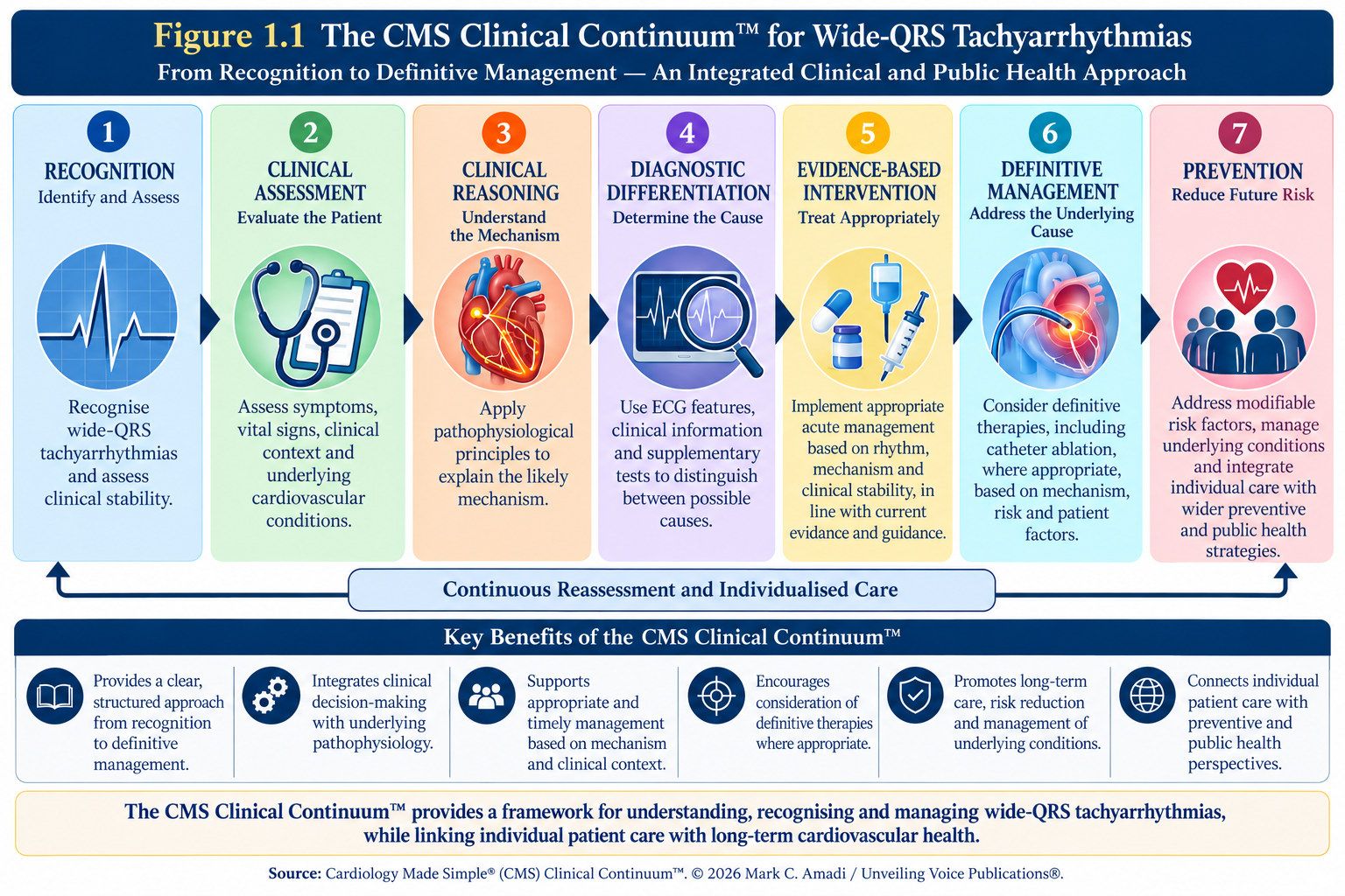
The CMS Clinical Continuum™ provides a conceptual overview of the wider clinical journey following recognition of a cardiac arrhythmia:
Recognition → Clinical Assessment → Clinical Reasoning → Diagnostic Differentiation → Evidence-Based Intervention → Definitive Management → Prevention
These stages represent interconnected domains of care rather than a mandatory sequence of bedside actions. In an acute presentation, assessment and intervention may need to occur immediately, before the precise arrhythmia mechanism has been established.1,2
Volume I introduces this broader framework while concentrating on recognition, physiological understanding and the foundations of clinical reasoning. Subsequent volumes will examine diagnostic differentiation, intervention and definitive management in greater detail
1.5 Defining Wide-QRS Tachyarrhythmia
A tachyarrhythmia is an abnormally rapid cardiac rhythm arising from altered impulse formation, impulse conduction or both. In adults, tachycardia is conventionally associated with a heart rate exceeding 100 beats per minute, although its clinical significance depends upon the rhythm mechanism and the patient’s physiological context.1
For the classification of tachycardias, a wide-QRS tachycardia is characterised by a QRS duration >120 milliseconds on the surface electrocardiogram (ECG); a narrow-QRS tachycardia has a QRS duration ≤120 milliseconds.1 This distinction describes the ECG appearance—it does not establish the underlying diagnosis.
QRS widening indicates altered ventricular activation. The impulse may originate within the ventricles, travel through an abnormal or delayed conduction pathway, or activate the ventricles in a sequence that differs from normal rapid conduction through the His–Purkinje system.1,2
Wide-QRS tachycardias may arise through several mechanisms, including:
ventricular tachycardia (VT);
supraventricular tachycardia with pre-existing bundle branch block or rate-related aberrancy;
tachycardia with antegrade conduction through an accessory atrioventricular pathway;
ventricular pacing; and
selected metabolic or pharmacological disturbances affecting ventricular conduction.1
From a practical electrophysiological perspective, the principal differential is between ventricular origin and supraventricular origin with abnormal ventricular activation.1,2 The ESC guideline reports that approximately 80% of presenting wide-QRS tachycardias are VT, around 15% represent SVT with bundle branch block or aberrant conduction, and approximately 5% involve antegrade conduction through an accessory pathway.1 These proportions provide useful clinical context but do not establish the diagnosis in an individual patient.
When the mechanism of a regular wide-QRS tachycardia remains uncertain, the rhythm should be approached cautiously and managed as VT until an alternative diagnosis is established.1
Recognising QRS widening identifies the abnormality; the next task is to determine the electrophysiological mechanism responsible for it. Clinical interpretation must therefore move beyond the descriptive question:
“Is the QRS wide?”
to the more clinically meaningful question:
“Why has ventricular activation become abnormal?”
1.6 Thinking Before Acting: The Discipline of Clinical Reasoning
Clinical reasoning begins with assessment of the patient and recognition of immediate risk. Haemodynamic instability, altered consciousness, ongoing ischaemic symptoms or other evidence of serious compromise may require urgent intervention before detailed rhythm differentiation is possible.1,2
When the patient is sufficiently stable, a 12-lead ECG, relevant clinical history and comparison with a previous ECG may help clarify the electrophysiological mechanism. Nevertheless, haemodynamic stability does not exclude ventricular tachycardia, and uncertainty concerning a regular wide-QRS tachycardia should not lead to inappropriate reassurance or delay in necessary treatment.1,2
The purpose of structured reasoning is to support safe clinical prioritisation while recognising the limitations of the available evidence.
Chapter Summary
Wide-QRS tachyarrhythmias require more than recognition of an abnormal ECG pattern. Similar appearances may arise from different electrophysiological mechanisms, making interpretation dependent upon integration of the ECG with the patient’s haemodynamic condition and clinical context.
A wide-QRS tachycardia is an electrocardiographic presentation, not a definitive diagnosis. Clinical reasoning must therefore move from recognition towards mechanism while prioritising immediate patient safety. When the mechanism of a regular wide-QRS tachycardia remains uncertain, it should be managed as VT until an alternative mechanism is established.1,2
The CMS Clinical Continuum™ places this reasoning within a broader pathway:
Recognition → Clinical Assessment → Clinical Reasoning → Diagnostic Differentiation → Evidence-Based Intervention → Definitive Management → Prevention
Together, these stages connect the presenting rhythm with immediate clinical priorities, mechanism-based reasoning, appropriate intervention, definitive management and longer-term cardiovascular prevention.
Key Take-Home Messages
A wide QRS is an observation, not a diagnosis. The underlying mechanism must still be determined.
Recognition begins the clinical process. ECG interpretation must be integrated with haemodynamic status, symptoms, cardiovascular history and relevant clinical findings.
Haemodynamic stability does not exclude ventricular tachycardia. When a regular wide-QRS tachycardia remains diagnostically uncertain, VT should remain the working diagnosis until an alternative mechanism is established.1,2
Clinical reasoning should move from description to mechanism. Ask where the impulse originated, which pathway it followed, how rapidly ventricular activation propagated and how the normal activation sequence changed.
Immediate priorities come before complete diagnostic certainty. Determine what requires action now and what can safely be clarified later.
Recognition may reveal more than the presenting arrhythmia. Structural heart disease, cardiomyopathy or inherited arrhythmogenic conditions may require subsequent specialist assessment, risk stratification or family evaluation.2,3
Arrhythmia care extends beyond the acute episode. The CMS Clinical Continuum™ connects recognition and intervention with definitive management and prevention.
References, Chapter 1
- 1. Brugada J, Katritsis DG, Arbelo E, et al. 2019 ESC Guidelines for the management of patients with supraventricular tachycardia. Eur Heart J. 2020;41(5):655–720. doi:10.1093/eurheartj/ehz467.
- 2. Zeppenfeld K, Tfelt-Hansen J, de Riva M, et al. 2022 ESC Guidelines for the management of patients with ventricular arrhythmias and the prevention of sudden cardiac death. Eur Heart J. 2022;43(40):3997–4126. doi:10.1093/eurheartj/ehac262.
- 3. Arbelo E, Protonotarios A, Gimeno JR, et al. 2023 ESC Guidelines for the management of cardiomyopathies. Eur Heart J. 2023;44(37):3503–3626. doi:10.1093/eurheartj/ehad194.
Chapter 2
Understanding Cardiac Electrophysiology
The Electrical Foundation of Wide-QRS Tachyarrhythmias
2.0 Introduction
Every heartbeat begins with an electrical impulse. Before the heart contracts and pumps blood, a coordinated sequence of electrical events occurs within its specialised conduction system. Electrical activation normally begins within the sinoatrial node, spreads through the atria to the atrioventricular node and subsequently enters the His–Purkinje system, where rapid conduction through the bundle branches and Purkinje network coordinates ventricular activation. This organised sequence enables electrical excitation to be translated into effective mechanical contraction.1
The electrocardiogram (ECG) provides a surface representation of this underlying electrical activity. Disturbances of impulse formation, conduction or both may produce cardiac arrhythmias, while abnormalities affecting the origin, pathway, speed or sequence of ventricular activation may alter QRS morphology and duration.1,2 Understanding normal cardiac electrophysiology is therefore fundamental to explaining how abnormal rhythms arise.
This is particularly important when approaching wide-QRS tachyarrhythmias. A wide QRS reflects altered ventricular activation, but understanding why activation has changed requires knowledge of how it normally occurs.
This chapter follows the normal electrical journey from impulse formation in the sinoatrial node, through atrial activation and the atrioventricular node, towards the specialised ventricular conduction system. Along the way, automaticity, excitability, conduction, depolarisation, repolarisation and refractoriness provide the physiological foundation for understanding normal cardiac rhythm and its abnormalities.
The emphasis throughout is physiology before pattern recognition. The ECG should not be viewed merely as a collection of waves and intervals to memorise, but as the surface expression of underlying electrical events.
2.1 The Heart’s Electrical Conduction System
The heart is not simply a muscular pump; it is an electromechanical organ whose coordinated function depends upon the generation and propagation of electrical impulses. During normal sinus rhythm, electrical activation follows an organised pathway through the specialised cardiac conduction system, allowing atrial activation to precede ventricular activation and supporting coordinated cardiac contraction.1
Unlike skeletal muscle, the heart possesses intrinsic electrical activity. Specialised pacemaker cells can generate spontaneous electrical impulses through automaticity, allowing cardiac electrical activity to continue without external neural stimulation. The autonomic nervous system modifies this intrinsic activity according to physiological demand.
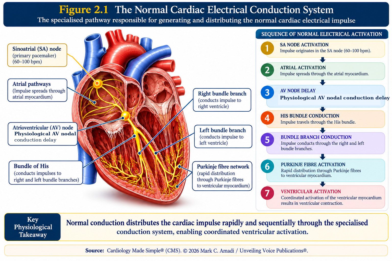
Normal electrical activation begins within the sinoatrial (SA) node, the heart’s principal physiological pacemaker. The impulse spreads through the atrial myocardium before reaching the atrioventricular (AV) node, where relatively slow conduction contributes to the normal temporal relationship between atrial and ventricular activation.
The impulse subsequently enters the bundle of His, which conducts activation into the specialised ventricular conduction system. The His bundle divides into the right and left bundle branches, from which activation is rapidly distributed through the Purkinje fibre network to the ventricular myocardium. This specialised pathway enables ventricular depolarisation to occur in an organised sequence.
The normal electrical pathway can therefore be represented as:
SA node → Atrial myocardium → AV node → His bundle → Right and left bundle branches → Purkinje network → Ventricular myocardium
This sequence is clinically important because QRS morphology and duration depend upon how the ventricles are activated. When activation proceeds rapidly through the specialised His–Purkinje system, ventricular depolarisation occurs in an organised sequence. When the origin, pathway, speed or sequence of activation changes, QRS morphology and duration may also change.
Figure 2.1 provides an overview of the specialised cardiac conduction system before its principal components are examined individually.
ECG clarification: The normal adult PR interval is approximately 120–200 ms, measured from the onset of the P wave to the onset of the QRS complex. It reflects atrial, AV junctional and His–Purkinje conduction, not isolated AV nodal delay.
2.2 The Sinoatrial Node: Where Every Normal Heartbeat Begins
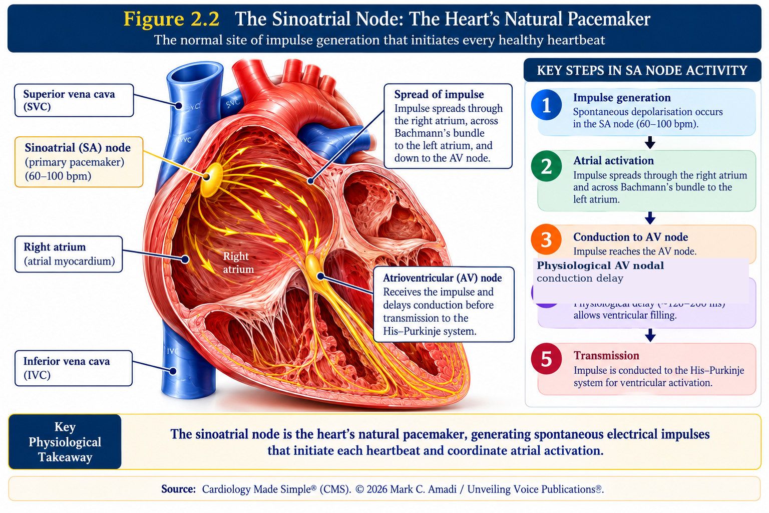
The sinoatrial (SA) node is the heart’s dominant physiological pacemaker and the normal site of impulse initiation. Located subepicardially in the superior right atrium near the junction of the superior vena cava and right atrium, it consists of specialised pacemaker cells within a complex architecture of nodal and surrounding atrial tissue. Under normal physiological conditions, the SA node establishes sinus rhythm because its spontaneous rate of impulse generation ordinarily exceeds that of subsidiary pacemaker tissues elsewhere in the conduction system.3,5
Unlike working atrial and ventricular myocytes, SA nodal pacemaker cells do not maintain a stable resting membrane potential. Instead, they undergo spontaneous diastolic depolarisation during phase 4, progressively approaching threshold and enabling rhythmic impulse generation without external neural stimulation. This pacemaker activity reflects the coordinated interaction of several ionic mechanisms, including the hyperpolarisation-activated funny current (I_f), calcium currents, potassium conductance and intracellular calcium cycling.3,6 Together, these mechanisms produce automaticity—the intrinsic capacity of pacemaker tissue to generate electrical impulses spontaneously.
Although the heart possesses intrinsic rhythmicity, the autonomic nervous system continuously modifies SA nodal activity according to physiological demand. Sympathetic stimulation, acting predominantly through β₁-adrenergic signalling, accelerates spontaneous diastolic depolarisation and increases heart rate. Parasympathetic stimulation, principally through vagal influences, slows pacemaker activity and reduces heart rate. These opposing influences allow sinus rate to adapt dynamically to exercise, rest, physiological stress and changing metabolic requirements.3,6
Once generated, the impulse spreads from the SA nodal region into the surrounding atrial myocardium, initiating organised atrial depolarisation. This electrical activity contributes to the P wave recorded on the surface ECG before activation progresses towards the atrioventricular node.1,3
If normal sinus impulse formation or transmission fails, subsidiary pacemaker tissues may generate escape rhythms. This hierarchical organisation provides a physiological safeguard, allowing cardiac electrical activity to continue when the dominant pacemaker or its transmission to surrounding atrial tissue is impaired.1,3
Understanding SA nodal physiology also helps explain abnormalities of sinus rhythm. Disturbances of impulse formation or sinoatrial conduction may produce sinus bradycardia, sinus pauses or arrest, sinoatrial exit block and other manifestations of sinus node dysfunction. Conversely, increased sinus automaticity may produce sinus tachycardia, while impulses originating outside the SA node may generate ectopic atrial rhythms.2,5,7
The SA node therefore provides the logical starting point for cardiac electrophysiology: automaticity generates the impulse, physiological regulation modifies its rate, and atrial conduction begins the organised electrical sequence that ultimately activates the ventricles.
Figure 2.2 illustrates the anatomical position of the SA node and its role as the starting point of normal cardiac electrical activation.
ECG clarification: The normal adult PR interval is approximately 120–200 ms, measured from the onset of the P wave to the onset of the QRS complex. It reflects atrial, AV junctional and His–Purkinje conduction, not isolated AV nodal delay.
2.3 The Atrioventricular Node: The Heart’s Physiological Gatekeeper
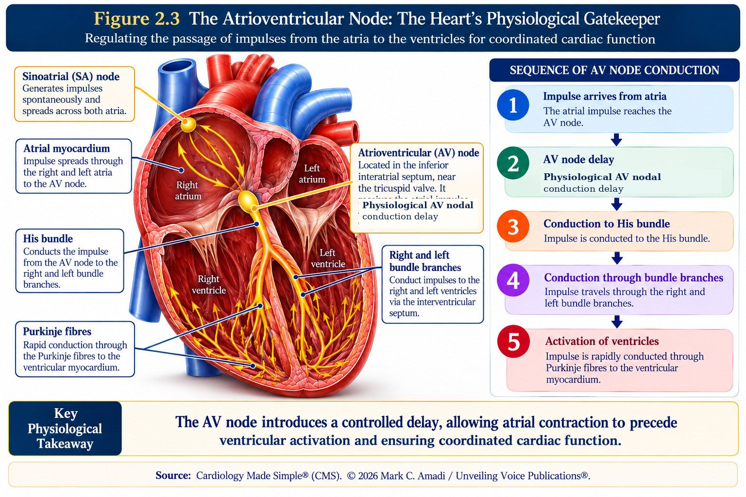
The atrioventricular (AV) node occupies a strategically important position between atrial and ventricular electrical activation. Located within the inferior interatrial septal region as part of the atrioventricular conduction axis, it receives electrical activation from the atria and conducts it towards the His bundle and specialised ventricular conduction system.3,4
Unlike the rapidly conducting His–Purkinje system, conduction through the AV nodal region is relatively slow. This physiological delay contributes to the normal temporal separation between atrial and ventricular activation, allowing atrial activation and contraction to precede ventricular activation.1,3
AV nodal conduction is influenced by the electrophysiological properties of nodal cells, including relatively slow calcium-dependent depolarisation, cellular architecture and decremental conduction. As atrial impulses arrive more rapidly, AV nodal conduction may progressively slow and some impulses may fail to conduct. Together with AV nodal refractoriness, this behaviour contributes to the node’s important role as a physiological filter between atrial and ventricular electrical activity.2,3 This filtering function becomes particularly important during rapid atrial rhythms. The AV node does not ordinarily transmit every rapidly arriving atrial impulse to the ventricles, helping to limit the ventricular response to excessive atrial electrical activity.2
The autonomic nervous system further modifies AV nodal behaviour. Sympathetic stimulation generally facilitates AV nodal conduction and shortens refractoriness, whereas parasympathetic stimulation slows conduction and increases refractoriness. These effects allow atrioventricular conduction to adapt to changing physiological conditions.3
On the surface ECG, atrioventricular conduction contributes to the PR interval. Importantly, the PR interval extends from the onset of atrial depolarisation to the onset of ventricular depolarisation and therefore reflects conduction through several structures; it should not be interpreted as a direct measurement of AV nodal conduction alone.1,4
The atrioventricular junction also contains tissues capable of subsidiary pacemaker activity. If higher pacemaker activity fails or is not successfully transmitted, junctional escape activity may provide a backup mechanism. It is therefore more precise to attribute this subsidiary automaticity to the AV junctional tissues rather than to the compact AV node alone.3,4
Abnormalities of atrioventricular conduction may manifest as first-degree, second-degree, high-grade or complete AV block. The anatomical site of conduction disturbance may lie within the AV node or below it within the His–Purkinje system and cannot always be determined from the PR interval alone.4,7
The AV nodal region is also important in atrioventricular nodal re-entrant tachycardia (AVNRT), in which functionally distinct pathways within or around the AV nodal region can participate in a re-entrant circuit.2 Understanding normal AV nodal physiology therefore provides a foundation for understanding both conduction disorders and selected supraventricular tachyarrhythmias.
A particularly important contrast occurs when an accessory atrioventricular pathway provides an alternative route between atrial and ventricular myocardium. An accessory pathway capable of antegrade conduction may allow electrical activation to bypass the normal AV nodal delay and filtering properties, producing ventricular pre-excitation. Its conduction properties influence the risk of rapid ventricular activation during atrial fibrillation.2
The AV node therefore performs more than a relay function. Through controlled delay, decremental conduction and refractoriness, it helps coordinate atrioventricular activation while regulating the transmission of atrial electrical activity towards the ventricles.
Figure 2.3 summarises the anatomical position of the AV node and its roles in physiological delay and regulation of atrioventricular conduction.
ECG clarification: The normal adult PR interval is approximately 120–200 ms, measured from the onset of the P wave to the onset of the QRS complex. It reflects atrial, AV junctional and His–Purkinje conduction, not isolated AV nodal delay.
Chapter Summary
Normal cardiac rhythm depends upon the coordinated generation and conduction of electrical impulses through the specialised cardiac conduction system. Electrical activation begins within the sinoatrial (SA) node, spreads through the atrial myocardium to the atrioventricular (AV) node and subsequently enters the His–Purkinje system before activating the ventricular myocardium.1,3
The SA node establishes the normal starting point of cardiac activation through automaticity, while autonomic influences modify its rate according to physiological demand.3,6 The AV node contributes controlled conduction delay, decremental conduction and refractoriness, helping to coordinate atrioventricular activation and regulate the transmission of atrial impulses towards the ventricles.2,3
These physiological principles provide the foundation for ECG interpretation. The ECG is the surface expression of underlying electrical events; understanding normal impulse formation and conduction therefore provides the reference point for recognising when cardiac activation becomes abnormal.
Key Take-Home Messages
The heart is an electromechanical organ. Coordinated contraction depends upon the organised generation and propagation of electrical impulses through the specialised conduction system.
The SA node is the dominant physiological pacemaker. Spontaneous phase 4 depolarisation enables automaticity, while autonomic influences modify sinus rate according to physiological demand.3,6
The origin of an impulse matters. Normal sinus activation begins in the SA node and spreads through the atria before reaching the AV node and specialised ventricular conduction system.
The AV node is a physiological gatekeeper. Controlled delay, decremental conduction and refractoriness help coordinate atrioventricular activation and ordinarily limit transmission of excessively rapid atrial activity.2,3
The PR interval is not a direct measurement of AV nodal conduction alone. It reflects atrioventricular conduction through several structures, and prolongation does not by itself establish the anatomical site of delay.1,4,7
Accessory pathways provide an important physiological contrast to normal AV nodal conduction. An accessory pathway capable of antegrade conduction may bypass normal AV nodal delay and filtering properties, producing ventricular pre-excitation.2
Interpret the ECG through physiology. Ask where the impulse originated, which pathway it followed, how rapidly it was conducted and in what sequence the ventricles were activated.
Normal physiology provides the reference point for recognising abnormality. Understanding normal impulse formation and conduction establishes the foundation for interpreting altered ventricular activation and wide-QRS tachyarrhythmias.
References, Chapter 2
- 1. Kennedy A, Finlay DD, Guldenring D, Bond R, Moran K, McLaughlin J. The cardiac conduction system: generation and conduction of the cardiac impulse. Crit Care Nurs Clin North Am. 2016;28(3):269–279. doi:10.1016/j.cnc.2016.04.001.
- 2. Brugada J, Katritsis DG, Arbelo E, et al. 2019 ESC Guidelines for the management of patients with supraventricular tachycardia. Eur Heart J. 2020;41(5):655–720. doi:10.1093/eurheartj/ehz467.
- 3. Park DS, Fishman GI. The cardiac conduction system: development, function and therapeutic targets. Nat Rev Cardiol. 2026;23(5):303–323. doi:10.1038/s41569-025-01227-x.
- 4. Karki R, Raina A, Ezzeddine FM, Bois MC, Asirvatham SJ. Anatomy and pathology of the cardiac conduction system. Cardiol Clin. 2023;41(3):277–292. doi:10.1016/j.ccl.2023.03.016.
- 5. Kalyanasundaram A, Li N, Augostini RS, Weiss R, Hummel JD, Fedorov VV. Three-dimensional functional anatomy of the human sinoatrial node for epicardial and endocardial mapping and ablation. Heart Rhythm. 2023;20(1):122–133. doi:10.1016/j.hrthm.2022.08.039.
- 6. Peters CH, Rickert C, Morotti S, et al. The funny current I_f is essential for the fight-or-flight response in cardiac pacemaker cells. J Gen Physiol. 2022;154(12):e202213193. doi:10.1085/jgp.202213193.
- 7. Glikson M, Nielsen JC, Kronborg MB, et al. 2021 ESC Guidelines on cardiac pacing and cardiac resynchronization therapy. Eur Heart J. 2021;42(35):3427–3520. doi:10.1093/eurheartj/ehab364.
Chapter 3
The His–Purkinje System and Ventricular Activation
The Physiological Basis of the Normal QRS Complex
From AV Nodal Delay to Rapid Ventricular Activation
Having traversed the atrioventricular (AV) conduction axis, the electrical impulse enters the specialised ventricular conduction system, where an important physiological transition occurs. Whereas conduction through the AV nodal region is relatively slow, the His–Purkinje system is specialised for rapid impulse transmission. This transition from controlled atrioventricular delay to rapid ventricular conduction enables the ventricles to be activated through an organised and coordinated sequence.1,2
The His–Purkinje system comprises the bundle of His, right and left bundle branches, their fascicular subdivisions and the Purkinje fibre network. Together, these specialised conducting tissues distribute electrical activation rapidly throughout the ventricles, allowing ventricular depolarisation to proceed through an organised spatial and temporal sequence over a relatively short interval.1,2
This rapid distribution of electrical activation is fundamental to the formation of the normal QRS complex. When ventricular activation proceeds through the specialised conduction system in the usual sequence, depolarisation is completed rapidly and the resulting QRS complex is generally narrow. QRS morphology and duration therefore provide important information about how the ventricles are being activated.1,2
When the origin, pathway, speed or sequence of ventricular activation changes, QRS morphology and duration may also change. Conduction delay or block within the bundle branches or fascicles may delay activation of part of the ventricular myocardium. Ventricular-origin impulses may begin outside the normal supraventricular activation sequence; ventricular pre-excitation may introduce activation through an accessory atrioventricular pathway; and ventricular pacing may alter the site and sequence from which ventricular depolarisation begins.2–4
Although these mechanisms differ electrophysiologically, they share an important principle: each alters the normal process of ventricular activation and may therefore alter the QRS complex.
This chapter follows the electrical impulse through the bundle of His, bundle branches and Purkinje fibre network before examining how their coordinated activity produces normal ventricular depolarisation. From this physiological foundation, the chapter explains why the normal QRS is generally narrow, how alterations in ventricular activation may produce QRS widening and why understanding these mechanisms is essential to the interpretation of wide-QRS tachyarrhythmias.
3.2 The Bundle of His: The Normal Electrical Bridge Between the Atria and Ventricles
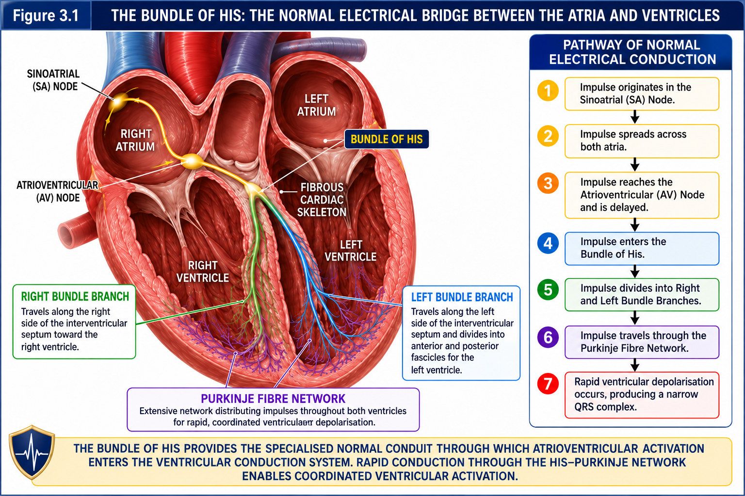
Following physiological slowing within the atrioventricular (AV) nodal region, the electrical impulse enters the bundle of His, marking its transition into the specialised ventricular conduction system. The His bundle is a compact tract of specialised conducting tissue that penetrates the fibrous atrioventricular junction before continuing along the upper interventricular septum. As the penetrating component of the normal AV conduction axis, it provides the specialised electrical pathway through which atrial activation normally gains access to the ventricular conduction system.1,2
The fibrous cardiac skeleton electrically insulates the working atrial myocardium from the working ventricular myocardium, preventing direct myocardial propagation between them under normal circumstances. Electrical activation therefore normally reaches the ventricular conduction system through the specialised AV conduction axis and penetrating His bundle. This anatomical organisation allows the physiological delay occurring predominantly within the AV nodal region to be followed by rapid transmission into the ventricular conduction system.1,2
Accessory atrioventricular pathways represent an important exception to this normal anatomical arrangement. These abnormal connections may provide an alternative route for electrical activation between atrial and ventricular myocardium, bypassing the normal AV nodal–His conduction pathway. When an accessory pathway conducts antegradely, it may alter the normal sequence of ventricular activation and produce ventricular pre-excitation.3
Unlike the AV nodal region, which is specialised for relatively slow and decremental conduction, the His–Purkinje system is adapted for rapid impulse transmission. Once the impulse enters the His bundle, it travels towards the right and left bundle branches and is subsequently distributed through the specialised ventricular conduction network.1,2
The His bundle therefore occupies a pivotal position within normal cardiac conduction: it forms the transition between controlled atrioventricular conduction and rapid ventricular activation. Disease affecting the His bundle may impair atrioventricular conduction, while abnormalities involving the more distal His–Purkinje system may produce fascicular conduction disturbances, bundle branch block or other forms of intraventricular conduction delay.4
Clinical safety qualification: Regular and irregular wide-QRS tachycardias require distinct assessment. An irregular wide-complex rhythm raises different diagnostic and medication-safety considerations, including the possibility of pre-excited atrial fibrillation. AV nodal-blocking drugs must be avoided in pre-excited atrial fibrillation; urgent management should follow current resuscitation and arrhythmia guidance.
3.3 The Right and Left Bundle Branches: Coordinating Ventricular Activation
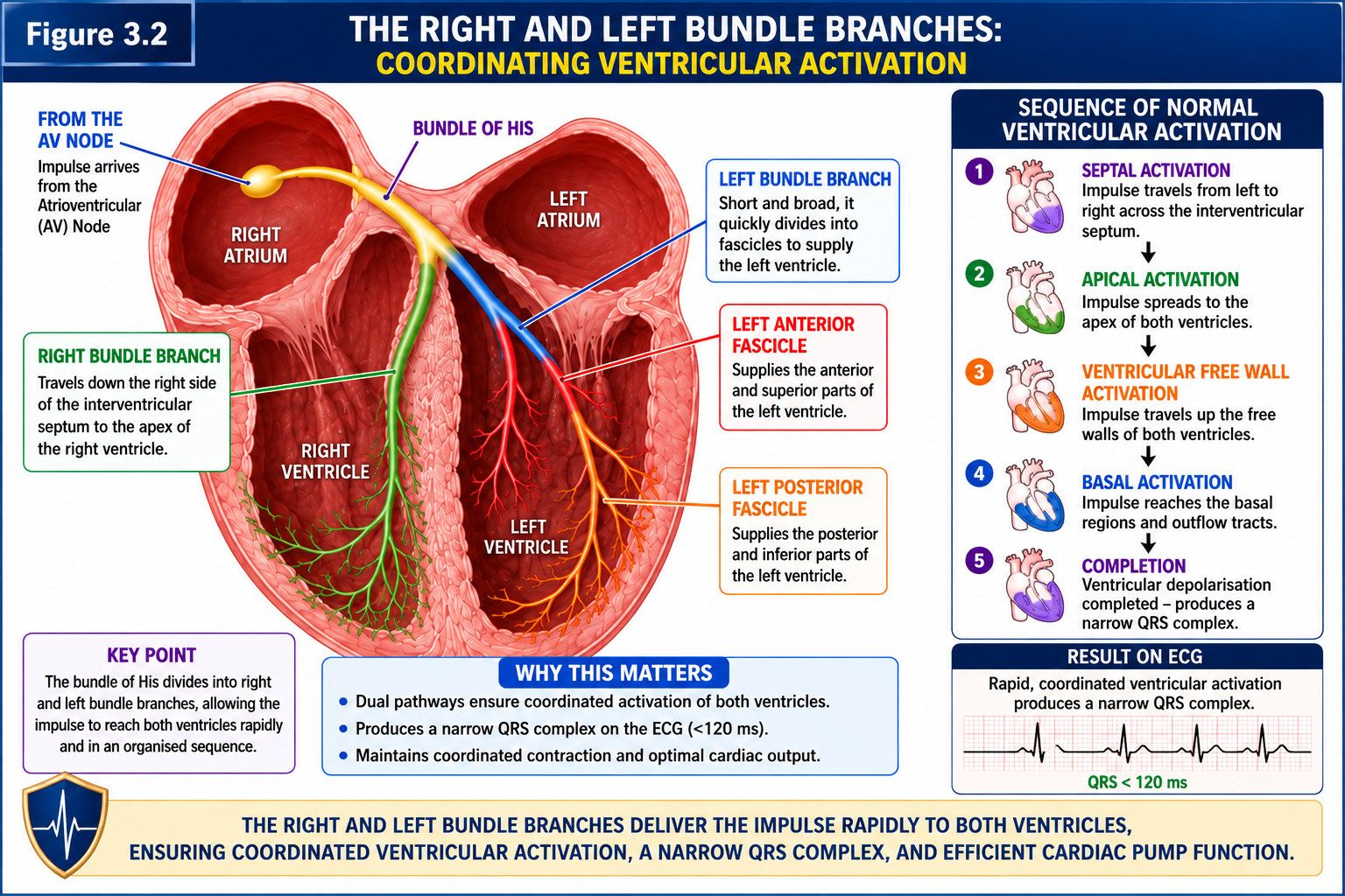
Following conduction through the bundle of His, the electrical impulse enters the right and left bundle branches, which distribute activation towards their respective ventricular territories. These specialised pathways form an integral part of the His–Purkinje system, enabling electrical activation to reach widely separated regions of the ventricles rapidly and in an organised sequence.1,2
The right bundle branch continues along the right side of the interventricular septum towards the right ventricle, where activation is distributed through the distal Purkinje network. The left bundle branch supplies the left ventricle through a more complex branching architecture, including major fascicular pathways that distribute activation across the left ventricular conduction system.1,2
This branching organisation is important because normal ventricular depolarisation does not occur everywhere at precisely the same instant. Rather, activation follows a rapid, organised spatial and temporal sequence. The bundle branches, together with the distal Purkinje network, allow this sequence to be completed efficiently over a relatively short interval.1,2
When conduction through a bundle branch or fascicular pathway is delayed or interrupted, the affected ventricular region may be activated later through alternative routes, including slower propagation through the working ventricular myocardium. The normal sequence of ventricular depolarisation therefore changes, potentially altering QRS morphology, electrical axis and duration.2,4 This provides the electrophysiological basis for bundle branch block and fascicular conduction abnormalities.
In right bundle branch block, right ventricular activation is delayed relative to the normal sequence. In left bundle branch block, left ventricular activation is substantially altered because rapid conduction through the normal left-sided His–Purkinje pathways is disrupted.2,4
Importantly, QRS widening does not establish bundle branch block as the mechanism. A wide QRS complex may also arise from ventricular-origin activation, ventricular pre-excitation, ventricular pacing or other disturbances that alter the origin, pathway, speed or sequence of ventricular activation.3,4
Figure 3.2 illustrates the right and left bundle branches and their coordinated contribution to rapid ventricular activation.
3.4 The Purkinje Fibre Network: The Heart’s High-Speed Ventricular Distribution System
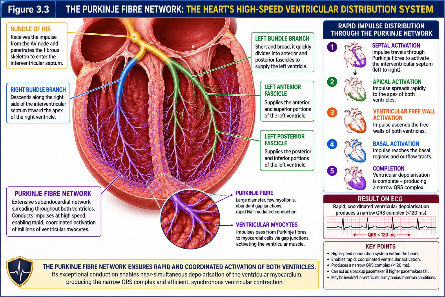
The Purkinje fibre network forms the distal component of the specialised ventricular conduction system. Arising from the terminal ramifications of the right and left bundle branches, Purkinje fibres form an extensive, predominantly subendocardial network that distributes electrical activation rapidly to widely separated regions of the ventricular myocardium.1,2
Purkinje fibres are among the fastest-conducting specialised tissues in the heart. Their cellular and electrophysiological properties support rapid impulse propagation, allowing electrical activation to reach multiple ventricular regions efficiently before spreading through the surrounding working myocardium. This high-speed distribution is fundamental to the organised sequence of ventricular depolarisation.1,2
Ventricular activation does not occur everywhere simultaneously. Depolarisation follows a coordinated spatial and temporal sequence. Early activation involves the interventricular septum, followed by activation of apical and ventricular free-wall regions, with later activation involving more basal regions. The precise sequence varies across ventricular regions and depends upon the distribution of the specialised conduction network.1,2 The Purkinje system rapidly distributes excitation across the ventricles, while propagation through the working ventricular myocardium completes the depolarisation process.
The electrical forces generated during this sequence contribute to the QRS complex recorded on the surface ECG. When rapid distribution through the normal His–Purkinje system is interrupted, bypassed or recruited abnormally, the sequence and duration of ventricular depolarisation may change, producing corresponding alterations in QRS morphology and duration.1,2
Purkinje tissue also possesses latent automaticity and may contribute to ventricular escape activity when higher pacemakers fail or when supraventricular impulses cannot reach the ventricles. This provides a physiological backup mechanism, although the emergence of a distal escape rhythm may indicate failure of higher-order impulse formation or interruption of conduction to the ventricles.1,4
Beyond its role in normal conduction, Purkinje tissue may participate directly in selected ventricular arrhythmias. Abnormal impulse formation or re-entrant mechanisms involving the Purkinje system can contribute to ventricular ectopy and ventricular tachyarrhythmias in particular clinical substrates. In selected patients, Purkinje-related triggers or circuits may be identified during electrophysiological evaluation and become targets for catheter ablation.5
The Purkinje network therefore completes the specialised pathway through which electrical activation is rapidly distributed across the ventricles. Its principal function is to deliver excitation efficiently to widely separated ventricular regions, enabling depolarisation to proceed through an organised and coordinated sequence.
Figure 3.3 illustrates the extensive Purkinje fibre network and its role in the rapid distribution of ventricular electrical activation.
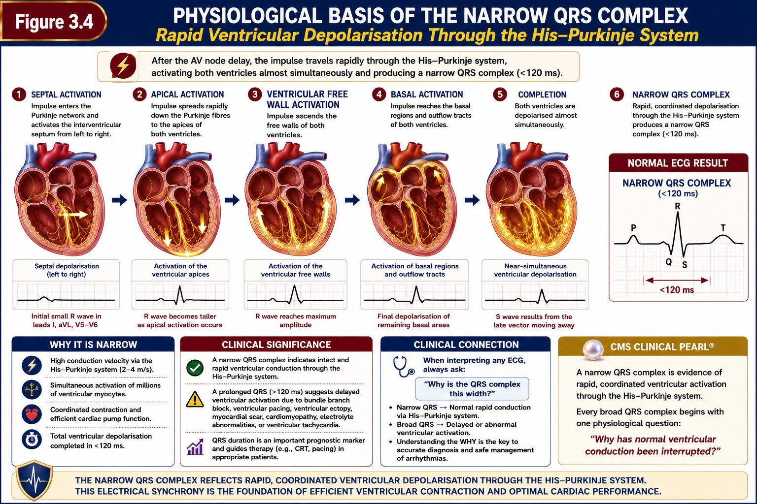
3.5 Why the Normal QRS Complex Is Narrow: Rapid Ventricular Depolarisation Explained
The QRS complex represents ventricular depolarisation as recorded on the surface electrocardiogram (ECG). Its morphology and duration reflect the origin, pathway, speed and sequence through which electrical activation spreads across the ventricles. Understanding why the QRS complex is normally narrow therefore requires understanding how ventricular activation is organised.1,2
Under normal conditions, the electrical impulse enters the ventricular conduction system through the bundle of His, divides into the right and left bundle branches and is distributed rapidly through the Purkinje fibre network. This specialised system delivers electrical excitation efficiently to widely separated ventricular regions, after which activation spreads through the working ventricular myocardium.1,2
Ventricular depolarisation does not occur everywhere simultaneously. Instead, it follows an organised spatial and temporal sequence. Early activation involves the interventricular septum, normally producing a predominantly left-to-right septal activation vector. Activation then progresses rapidly towards apical and ventricular free-wall regions, with later activation involving more basal regions. The electrical forces generated during this sequence combine to produce the QRS complex recorded on the surface ECG.1,2
The QRS is therefore narrow not because all ventricular myocytes depolarise at precisely the same instant, but because the His–Purkinje system distributes electrical activation rapidly and efficiently, allowing overall ventricular depolarisation to be completed within a relatively short interval.1,2
An important distinction is required when describing QRS duration. In the ESC classification of tachycardias, a QRS duration of ≤120 milliseconds is classified as narrow, whereas a QRS duration of >120 milliseconds is classified as wide.3 This threshold is useful for the clinical classification of tachycardia, but it should not be interpreted as meaning that every QRS measuring 120 milliseconds represents physiologically normal ventricular conduction.
When ventricular activation no longer follows the usual rapid His–Purkinje sequence, QRS morphology and duration may change. This may occur when conduction is delayed within the specialised conduction system, when activation originates within the ventricular myocardium, when an accessory pathway alters the normal sequence of depolarisation, or when ventricular pacing changes the site from which activation begins.3,4
The underlying physiological principle can therefore be summarised:
QRS duration reflects the overall time required for ventricular depolarisation.
QRS morphology reflects the direction, timing and relative contributions of the electrical forces generated during that process.
A narrow QRS generally indicates rapid ventricular activation through, or with early recruitment of, the specialised His–Purkinje system. Conversely, a wide QRS indicates prolonged ventricular depolarisation, which may result from delayed conduction or an altered activation pathway or sequence. Importantly, QRS widening alone does not identify the underlying mechanism.3,4
Figure 3.4 illustrates how rapid, organised ventricular activation through the His–Purkinje system contributes to the formation of a narrow QRS complex.
3.6 When Ventricular Activation Becomes Abnormal: The Physiological Basis of the Wide QRS Complex
Normal ventricular depolarisation depends upon rapid and organised electrical activation through the His–Purkinje system. This specialised conduction network distributes the impulse efficiently across the ventricles, allowing ventricular depolarisation to be completed within a relatively short interval and generally producing a narrow QRS complex.1,2
Ventricular activation becomes abnormal when this normal process is disrupted or replaced. The disturbance may involve the origin of the impulse, the pathway through which it travels, the speed at which it propagates or the sequence in which ventricular regions are activated. The ventricles may consequently be activated later, more slowly, through an alternative route or in a different spatial and temporal sequence.
This explains the physiological basis of QRS widening. When ventricular depolarisation can no longer be completed through the usual rapid and organised activation sequence, the total time required for ventricular activation may increase. Prolongation of that process may produce a wider QRS complex, while changes in the direction and sequence of activation may alter QRS morphology.1,2
For the clinical classification of tachycardias, QRS duration provides an important distinction between narrow- and wide-QRS presentations. The threshold adopted in this handbook follows the classification specified in the cited ESC supraventricular tachycardia guideline and should be applied consistently throughout the text, figures and definitions.3
QRS duration reflects the time required for ventricular depolarisation. A prolonged QRS indicates delayed or altered ventricular activation but does not independently establish whether the rhythm originates within the ventricles or above them. Interpretation therefore requires consideration of the impulse’s origin, conduction pathway, propagation speed and ventricular activation sequence.
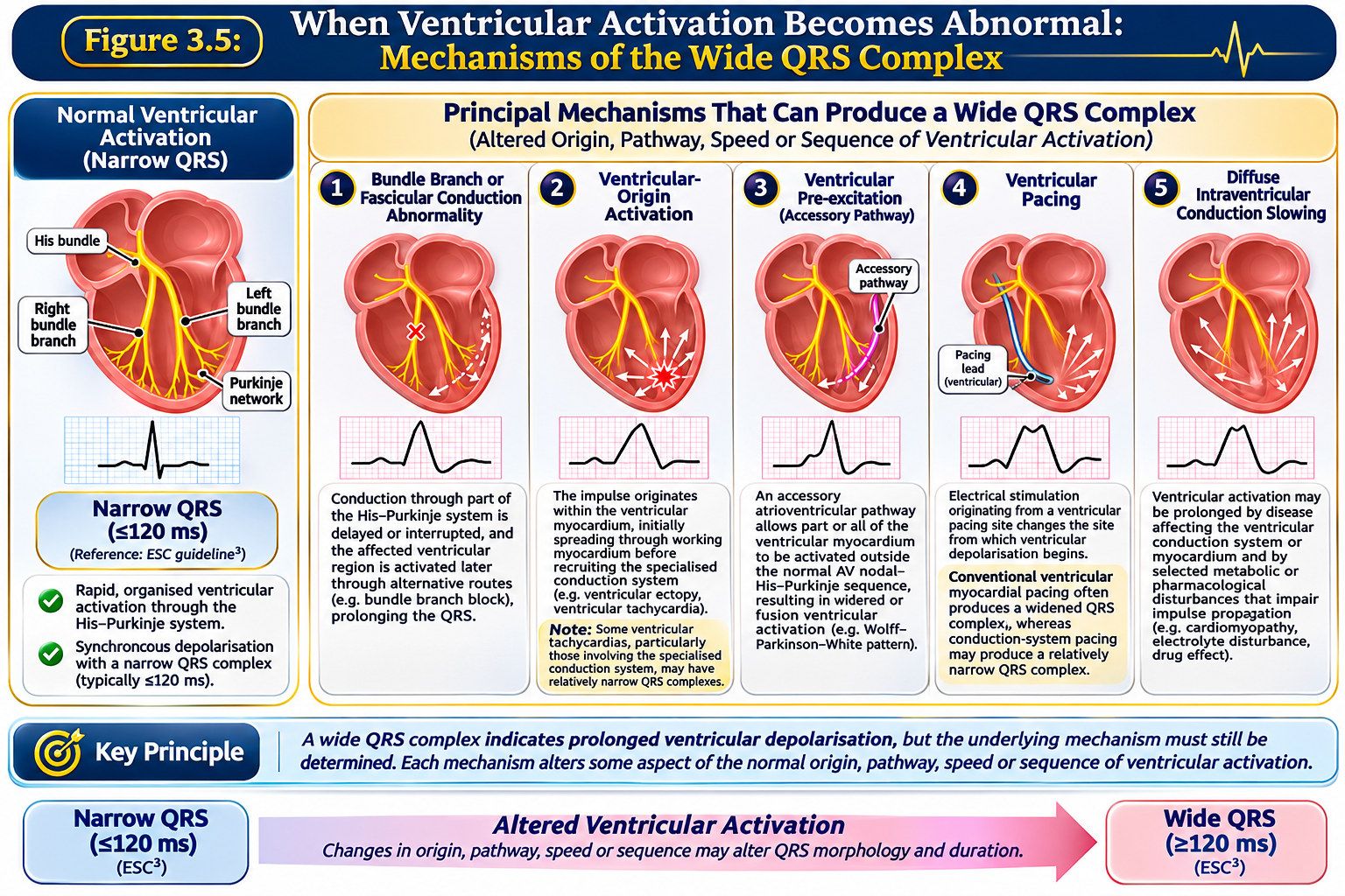
Several electrophysiological disturbances can produce QRS widening. Although they differ in anatomical location and mechanism, each alters some aspect of the normal origin, pathway, speed or sequence of ventricular activation. Five principal mechanisms provide a useful physiological framework for understanding how QRS widening may occur:
Bundle branch or fascicular conduction abnormality. When conduction through part of the His–Purkinje system is delayed or interrupted, the affected ventricular region may be activated later through alternative routes, including slower propagation through working ventricular myocardium. The normal sequence of depolarisation is therefore altered, potentially prolonging the QRS complex.2,4
Ventricular-origin activation. When an impulse originates within the ventricular myocardium rather than reaching the ventricles through the normal supraventricular conduction pathway, activation may initially spread through working myocardium before recruiting the specialised conduction system. Ventricular ectopy and ventricular tachycardia may therefore produce wide QRS complexes, with morphology influenced by the site and direction of ventricular activation. However, some ventricular tachycardias involving the specialised conduction system may produce relatively narrow QRS complexes.2,5
Ventricular pre-excitation. An accessory atrioventricular pathway capable of antegrade conduction may allow part or all of the ventricular myocardium to be activated outside the normal AV nodal–His–Purkinje sequence. The resulting alteration or fusion of ventricular activation through the accessory pathway and normal conduction system may change QRS morphology and duration.3
Ventricular pacing. Electrical stimulation originating from a ventricular pacing site changes the site from which ventricular depolarisation begins. Conventional ventricular myocardial pacing commonly produces an altered and widened QRS complex because activation follows a sequence different from normal intrinsic His–Purkinje conduction. However, conduction-system pacing may recruit the specialised conduction network and produce a relatively narrow QRS complex.4
Diffuse intraventricular conduction slowing. Ventricular activation may also be prolonged by disease affecting the ventricular conduction system or myocardium and by selected metabolic or pharmacological disturbances that impair impulse propagation. QRS widening arising in these settings should therefore be interpreted within the wider clinical context.2,4
These mechanisms demonstrate an important clinical principle: a wide QRS complex indicates prolonged ventricular depolarisation, but the underlying mechanism must still be determined. This requires integration of QRS morphology and duration with rhythm characteristics, atrial activity and the wider clinical context, including structural heart disease, medications, electrolyte abnormalities and other relevant findings.2–4 During tachycardia, ventricular tachycardia, supraventricular tachycardia with aberrant conduction and tachycardia involving ventricular pre-excitation may produce superficially similar wide-QRS appearances despite fundamentally different electrophysiological mechanisms.2,3 The clinical task is therefore to determine what has changed in ventricular activation and why.
Figure 3.5 brings these principles together by comparing normal ventricular activation with the principal mechanisms capable of producing QRS widening.
3.7 Clinical Integration: From Normal Ventricular Activation to Wide-QRS Tachyarrhythmias
Understanding normal ventricular activation provides the reference point for interpreting wide-QRS tachyarrhythmias. A wide QRS indicates prolonged ventricular depolarisation, but QRS width alone does not identify the underlying electrophysiological mechanism.1,2
This distinction becomes particularly important during tachycardia. Ventricular tachycardia, supraventricular tachycardia with aberrant conduction and tachycardia involving ventricular pre-excitation may produce similar wide-QRS appearances despite fundamentally different electrophysiological mechanisms.2,3 The clinical challenge is therefore to determine why ventricular depolarisation is prolonged, rather than relying on QRS duration alone.
The physiology developed throughout this chapter provides a structured approach:
ORIGIN → PATHWAY → SPEED → SEQUENCE
Where did the impulse originate?
Which pathway did it follow?
How rapidly did it propagate?
In what sequence were the ventricles activated?
These questions connect ventricular physiology with mechanism-based ECG interpretation. They direct attention towards whether normal His–Purkinje activation has been delayed, bypassed, initiated from a ventricular focus or otherwise altered.
The clinical reasoning pathway can therefore be expressed as:
Understand normal activation → Recognise altered activation → Identify what changed → Determine the likely mechanism → Integrate the ECG with the patient’s clinical context.
Physiological understanding does not eliminate diagnostic uncertainty. ECG interpretation must also consider rhythm characteristics, clinical presentation and relevant cardiovascular disease. The transition from a normal QRS to a wide-QRS tachyarrhythmia therefore requires an explanation of the underlying ventricular activation mechanism, rather than QRS measurement alone.2,3
Chapter Summary
This chapter examined the specialised ventricular conduction system responsible for the rapid and organised distribution of electrical activation throughout the ventricles. Beginning with the bundle of His, the electrical impulse was followed through the right and left bundle branches and Purkinje fibre network, demonstrating how the His–Purkinje system enables ventricular depolarisation to proceed through a coordinated spatial and temporal sequence.1,2
Rapid ventricular activation through this specialised network provides the physiological basis for the generally narrow QRS complex. QRS morphology and duration reflect the origin, pathway, speed and sequence of ventricular depolarisation.
When normal ventricular activation is disrupted or replaced, depolarisation may proceed more slowly, through an alternative pathway or from a different site, altering QRS morphology and potentially widening the QRS complex. Principal mechanisms include bundle branch or fascicular conduction abnormalities, ventricular-origin activation, ventricular pre-excitation, ventricular pacing and diffuse intraventricular conduction slowing.2–4
A wide QRS complex is therefore an electrical finding, not a diagnosis. Its interpretation requires integration of ventricular activation physiology with ECG morphology, rhythm characteristics and the wider clinical context.2,3
These principles establish the electrophysiological foundation for progressing from recognition of a wide QRS towards mechanism-based interpretation of wide-QRS tachyarrhythmias.
Key Take-Home Messages
The His–Purkinje system enables rapid and organised ventricular activation. The His bundle, bundle branches and Purkinje network distribute electrical excitation efficiently throughout the ventricles.1,2
Normal ventricular activation is coordinated, not literally simultaneous. Depolarisation follows an organised spatial and temporal sequence.1,2
The QRS complex is the surface electrical expression of ventricular depolarisation. Its morphology and duration reflect the origin, pathway, speed and sequence of ventricular activation.1,2
A narrow QRS generally reflects rapid ventricular activation through, or with early recruitment of, the specialised His–Purkinje system. Alterations in this process may change QRS morphology and duration.1,2
A wide QRS complex is an electrical finding, not a diagnosis. Principal mechanisms include bundle branch or fascicular conduction abnormalities, ventricular-origin activation, ventricular pre-excitation, ventricular pacing and diffuse intraventricular conduction slowing.2–4
QRS width alone cannot determine the underlying electrophysiological mechanism. Interpretation requires integration of QRS morphology and duration with rhythm characteristics and the wider clinical context.2,3
Four questions provide the central physiological framework for QRS interpretation: ORIGIN → PATHWAY → SPEED → SEQUENCE. Where did the impulse originate? Which pathway did it follow? How rapidly did it propagate? In what sequence were the ventricles activated?
References, Chapter 3
- 1. Park DS, Fishman GI. The cardiac conduction system: development, function and therapeutic targets. Nat Rev Cardiol. 2026;23(5):303–323. doi:10.1038/s41569-025-01227-x.
- 2. Karki R, Raina A, Ezzeddine FM, Bois MC, Asirvatham SJ. Anatomy and pathology of the cardiac conduction system. Cardiol Clin. 2023;41(3):277–292. doi:10.1016/j.ccl.2023.03.016.
- 3. Brugada J, Katritsis DG, Arbelo E, et al. 2019 ESC Guidelines for the management of patients with supraventricular tachycardia. Eur Heart J. 2020;41(5):655–720. doi:10.1093/eurheartj/ehz467.
- 4. Glikson M, Nielsen JC, Kronborg MB, et al. 2021 ESC Guidelines on cardiac pacing and cardiac resynchronization therapy. Eur Heart J. 2021;42(35):3427–3520. doi:10.1093/eurheartj/ehab364.
- 5. Nogami A, Komatsu Y, Talib AK, Phanthawimol W, Naeemah QJ, Haruna T, Morishima I. Purkinje-related ventricular tachycardia and ventricular fibrillation: solved and unsolved questions. JACC Clin Electrophysiol. 2023;9(10):2172–2196. doi:10.1016/j.jacep.2023.05.040.
Interlude
Beyond the ECG: From Clinical Recognition to Cardiovascular Prevention
A wide-QRS tachyarrhythmia may initially present as an individual clinical problem: a patient develops symptoms, an abnormal rhythm is identified on the electrocardiogram (ECG), haemodynamic status is assessed and appropriate treatment is initiated. However, the significance of that encounter may extend beyond the immediate rhythm disturbance. Viewed through a preventive perspective, clinical recognition can become the starting point for identifying and reducing future cardiovascular risk.
The immediate priority is safe assessment and management. Once the patient’s condition permits, attention should also turn to an important question: Why did this arrhythmia occur? Investigation of the underlying mechanism and substrate may identify structural heart disease, cardiomyopathy, an accessory atrioventricular pathway, an inherited arrhythmogenic condition or potentially modifiable factors requiring further assessment and management.1–3
This creates a bridge between acute cardiovascular care, prevention of arrhythmia recurrence and longer-term cardiovascular risk management. Restoration of a stable rhythm does not necessarily represent the end of the patient’s clinical journey. Depending on the mechanism, underlying disease and individual risk profile, subsequent care may include risk stratification, electrophysiological assessment, catheter ablation, optimisation of medical therapy, cardiac pacing, implantable cardioverter-defibrillator therapy, cardiovascular risk-factor management and structured follow-up.1–4
The implications may extend beyond the individual patient. When an inherited cardiomyopathy or arrhythmogenic disorder is suspected or confirmed, appropriate genetic counselling, genetic evaluation and family screening may identify relatives who require further assessment.2,3 Recognition in one patient can therefore create an opportunity for family-level risk identification and prevention.
There is also a health-system dimension. Safe arrhythmia care depends on clinicians recognising potentially dangerous rhythms, responding appropriately to haemodynamic instability, managing diagnostic uncertainty and ensuring continuity between acute, specialist and preventive services. Effective clinical education can support these processes by strengthening physiological understanding, clinical judgement and appropriate referral.
Clinical cardiology and cardiovascular prevention therefore converge around a common question: What happens after the immediate problem has been recognised and treated? Clinical assessment establishes the likely rhythm mechanism and guides appropriate management. The preventive perspective considers how recurrence and future cardiovascular complications may be reduced and whether underlying or inherited disease warrants further investigation.
These considerations support viewing arrhythmia care as a continuum rather than a series of isolated encounters. Recognition of the presenting rhythm may lead to acute assessment, diagnostic clarification, risk stratification, appropriate intervention and longer-term prevention. The sequence and extent of these activities depend on the patient’s condition and clinical findings.
This relationship is illustrated in Figure Interlude 1, through the CMS Clinical Continuum™, an educational framework connecting acute recognition with subsequent cardiovascular care.
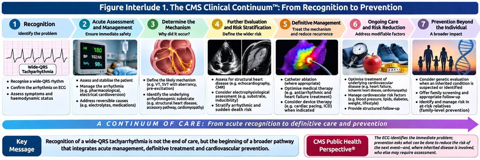
The continuum does not necessarily end when the rhythm terminates. It connects immediate clinical recognition with investigation, appropriate treatment and longer-term risk reduction. The ECG may therefore provide an entry point into a wider pathway of cardiovascular assessment, care and prevention.
Appendix A
CMS Quick Review®
Rapid Review of the Core Clinical and Electrophysiological Principles
The CMS Quick Review® consolidates the principal concepts developed throughout this volume into concise clinical review frameworks. It is designed for rapid revision and point-of-learning recall rather than as a substitute for the detailed physiological and clinical discussions within the chapters.
The reviews follow the central educational progression of Cardiology Made Simple®:
Understand the normal → Recognise what has changed → Explain the mechanism → Apply the reasoning clinically
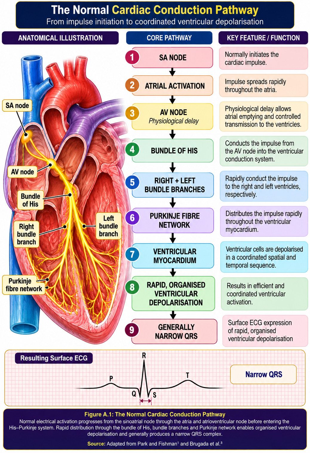
A.1 The Normal Cardiac Conduction Pathway
Purpose: Establish the normal electrical pathway against which abnormal ventricular activation and wide-QRS rhythms can be understood.
Normal cardiac activation follows an organised electrical journey from impulse initiation in the sinoatrial node to rapid ventricular depolarisation through the specialised His–Purkinje system.
Quick Clinical Interpretation
The normal conduction pathway provides the physiological reference point for ECG interpretation. The SA node initiates the impulse, the AV node delays its transmission, the His–Purkinje system rapidly distributes electrical activation through the ventricles, and the ventricular myocardium depolarises in an organised sequence, producing the QRS complex.
A.2 The Four Questions Behind the QRS
Purpose: Provide the central physiological framework for interpreting ventricular activation.
Every QRS complex reflects four fundamental properties of ventricular activation. Considering these properties helps move ECG interpretation from recognising a pattern towards understanding the mechanism responsible for it.
ORIGIN → PATHWAY → SPEED → SEQUENCE
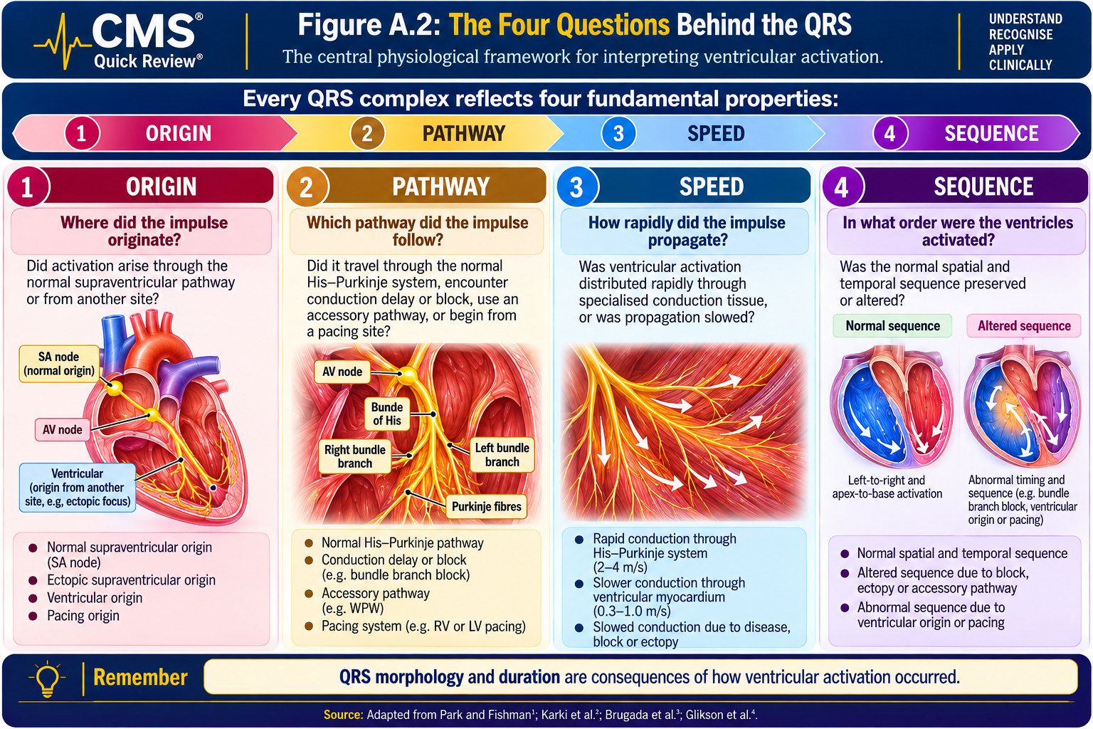
Quick Clinical Interpretation
ORIGIN: Where did the impulse originate?
PATHWAY: Which pathway did it follow?
SPEED: How rapidly did it propagate?
SEQUENCE: In what sequence were the ventricles activated?
Changes in one or more of these properties may alter QRS morphology or duration. The framework therefore provides a physiological starting point for investigating abnormal ventricular activation.
A.3 From Normal Ventricular Activation to the Wide QRS
Purpose: Explain how alterations in ventricular activation may produce QRS widening.
Normal and abnormal ventricular activation can be compared by examining the origin, pathway, speed and sequence of ventricular depolarisation. Changes in these properties may alter QRS morphology or duration, although an abnormal activation sequence does not invariably produce a wide QRS complex.
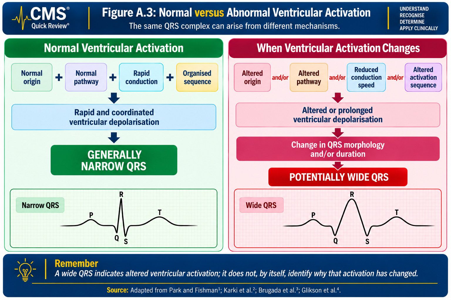
Quick Clinical Interpretation
A wide QRS indicates prolonged ventricular depolarisation. This may result from altered impulse origin, conduction pathway, propagation speed or activation sequence.
The key clinical question is therefore:
What has changed in ventricular activation to produce the wide QRS?
Understanding that change provides the physiological bridge between recognising QRS widening and investigating its underlying mechanism.
A.4 Five Principal Mechanisms of QRS Widening
Purpose: Connect QRS widening with the principal electrophysiological mechanisms capable of producing it.
Different electrophysiological disturbances may alter ventricular activation through distinct mechanisms yet produce a similar surface ECG finding: a wide QRS complex. Figure A.4 summarises five principal mechanisms.
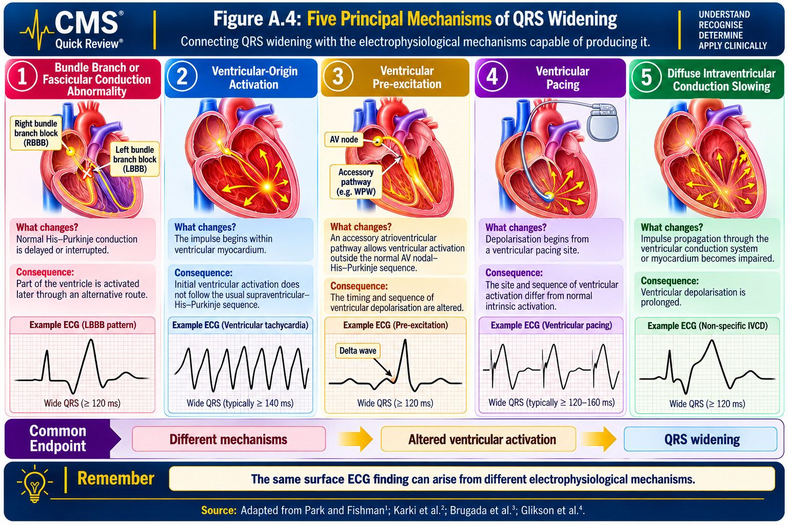
Quick Clinical Interpretation
Five principal mechanisms provide a useful physiological framework for understanding QRS widening:
Bundle branch or fascicular conduction abnormality: Delayed or interrupted conduction through the specialised ventricular conduction system may prolong ventricular depolarisation.
Ventricular-origin activation: Depolarisation begins within ventricular tissue rather than following the usual supraventricular activation sequence. Subsequent propagation may produce a wide QRS complex.
Ventricular pre-excitation: An accessory atrioventricular pathway capable of antegrade conduction activates part or all of the ventricular myocardium outside the normal AV nodal–His–Purkinje sequence.
Ventricular pacing: Ventricular activation initiated by a pacing stimulus may follow an altered sequence and produce QRS widening, depending on the pacing site and conduction-system recruitment.
Diffuse intraventricular conduction slowing: Ventricular depolarisation may be prolonged by myocardial or conduction-system disease, electrolyte disturbances or medications that impair impulse propagation.
Although these mechanisms differ, they share an important principle:
A wide QRS identifies prolonged ventricular depolarisation; it does not, by itself, establish the underlying mechanism.
A.5 Approaching the Wide-QRS Tachyarrhythmia
Purpose: Connect recognition of a wide-QRS tachyarrhythmia with immediate clinical assessment and structured physiological reasoning.
For the classification of tachycardias in this handbook, a wide-QRS tachycardia has a QRS duration greater than 120 ms, consistent with the convention used in the main chapters. QRS duration describes ventricular activation and does not independently establish the arrhythmia mechanism.
Recognising QRS widening is only the beginning. The immediate clinical priority is to assess the patient, identify haemodynamic instability and initiate appropriate emergency management when indicated. Mechanism-based ECG interpretation follows alongside clinical assessment and must not delay urgent treatment.
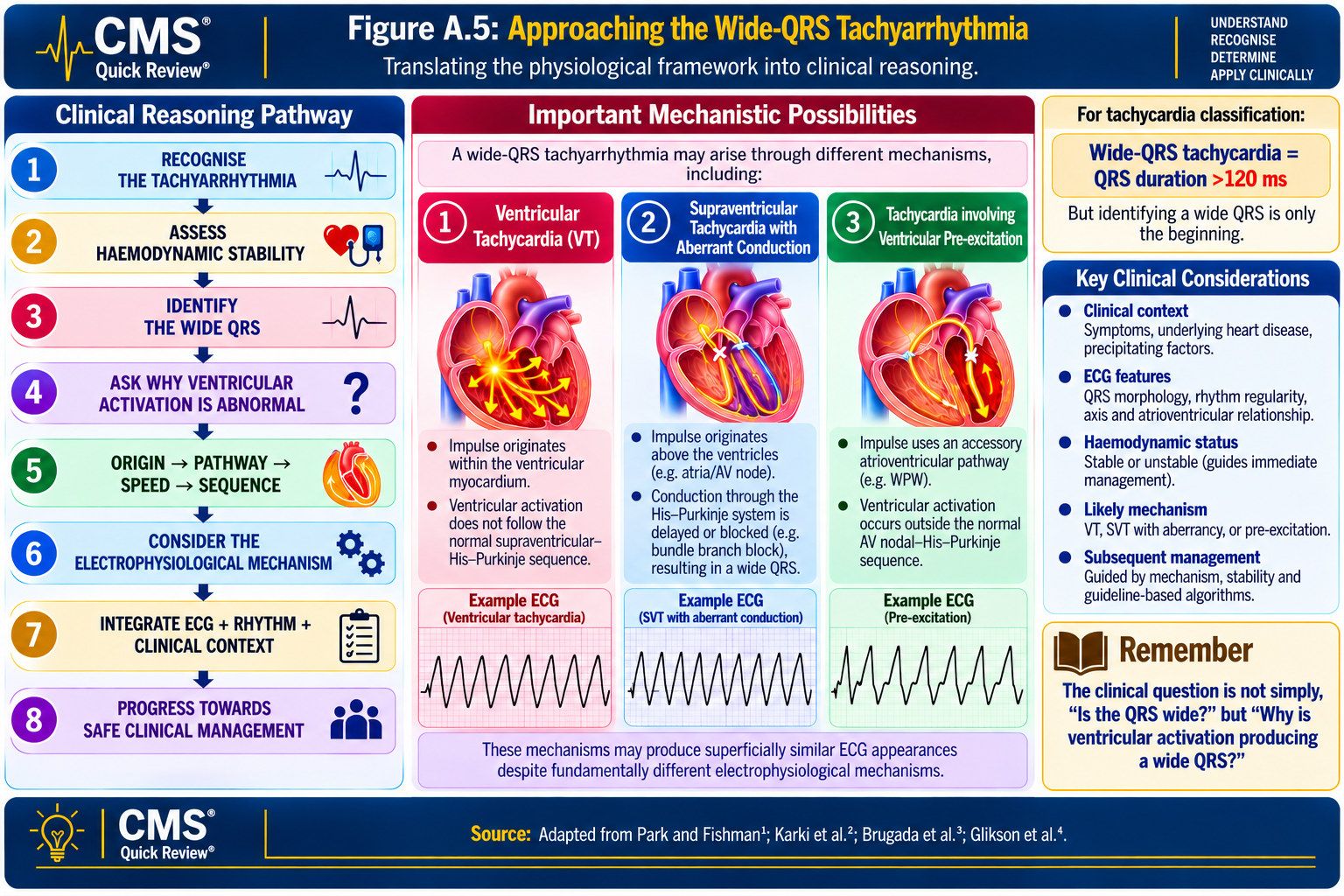
Quick Clinical Interpretation
When confronted with a wide-QRS tachyarrhythmia, apply the following clinical reasoning framework:
RECOGNISE: Is a wide-QRS tachycardia present?
ASSESS: Is the patient haemodynamically stable? Are there features requiring immediate intervention?
REASON: What has changed in the origin, pathway, speed or sequence of ventricular activation?
DIFFERENTIATE: Which electrophysiological mechanisms could explain the rhythm? If the mechanism remains uncertain, ventricular tachycardia must remain a primary consideration.
INTEGRATE: How do the ECG findings relate to the patient’s presentation, cardiovascular history and other clinical information?
Patient-safety principle: These questions support clinical reasoning; they do not replace emergency assessment or established resuscitation and arrhythmia-management guidance.
Do not stop at recognising a wide QRS. Assess the patient promptly, manage immediate risk and investigate the mechanism without delaying necessary treatment.
A.6 The CMS Clinical Continuum™
Purpose: Connect cardiovascular recognition with clinical assessment, diagnostic reasoning, appropriate treatment, longer-term management and prevention.
The CMS Clinical Continuum™ presents cardiovascular care as a connected process extending from recognition of the immediate problem to investigation, treatment and prevention. It is an author-developed educational framework rather than a validated clinical algorithm.
RECOGNITION → CLINICAL ASSESSMENT → CLINICAL REASONING → DIAGNOSTIC DIFFERENTIATION → EVIDENCE-BASED INTERVENTION → DEFINITIVE MANAGEMENT → PREVENTION
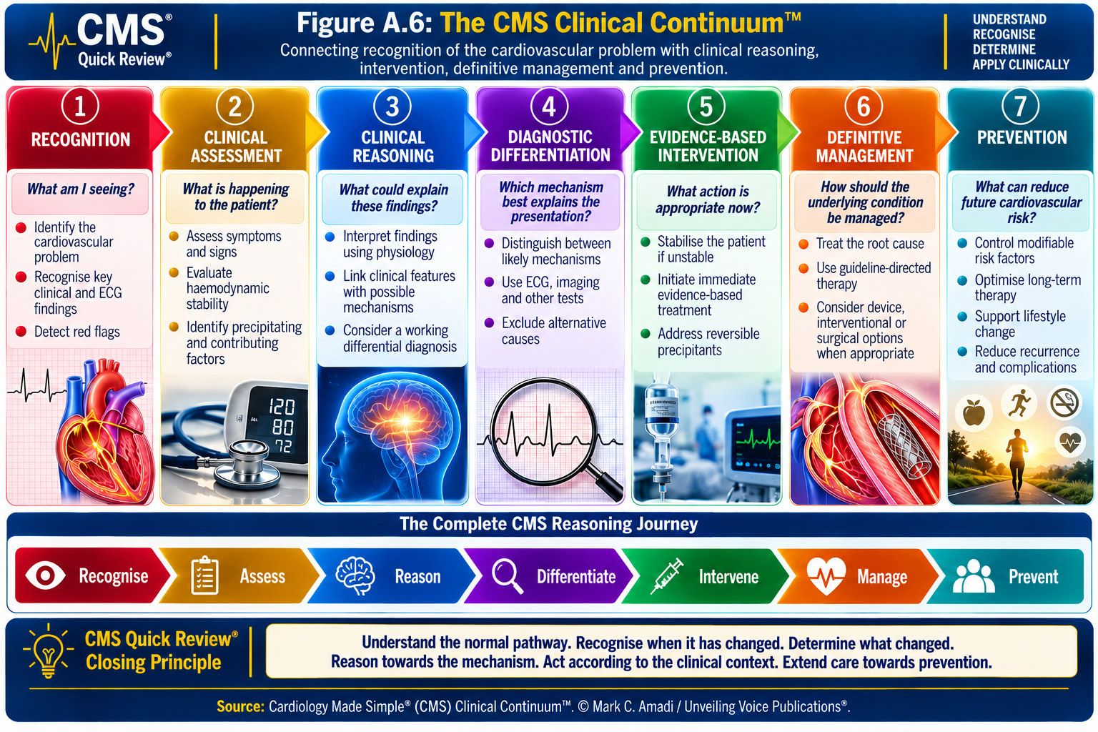
Quick Clinical Interpretation
The continuum encourages consideration of the wider patient journey:
Recognition: Identifies the presenting cardiovascular problem.
Clinical Assessment: Establishes the patient’s immediate clinical condition and haemodynamic status.
Clinical Reasoning: Integrates physiological understanding, ECG findings and clinical information.
Diagnostic Differentiation: Evaluates the likely underlying electrophysiological mechanism.
Evidence-Based Intervention: Addresses the immediate clinical problem according to its urgency and applicable guidance.
Definitive Management: Addresses the underlying arrhythmia mechanism or cardiovascular substrate when indicated.
Prevention: Considers how recurrent arrhythmia, future cardiovascular complications and relevant inherited risks may be reduced.
These components are interconnected and do not necessarily occur in a fixed chronological order. Urgent intervention may be required before definitive diagnostic clarification, while preventive assessment may begin during ongoing treatment.
The Central Principle Is:
Recognition begins the clinical journey; prevention completes the continuum of care.
Source: Cardiology Made Simple® (CMS) Clinical Continuum™. © Mark C. Amadi / Unveiling Voice Publications®.
Appendix B
CMS Learning Framework™
From Understanding to Clinical Application
The CMS Learning Framework™ defines the educational approach underpinning Cardiology Made Simple®. It is founded on the principle that effective clinical learning requires more than memorising diagnostic criteria, ECG patterns or treatment algorithms. Clinical knowledge becomes meaningful when the learner understands the underlying physiology, recognises when that physiology has changed, reasons towards the likely mechanism and applies evidence appropriately within the patient’s clinical context.
The framework connects scientific understanding with clinical reasoning and application, encouraging learners to progress from recognising what is happening, to understanding why it is happening, and determining what should happen next.
Four principles underpin this approach.
B.1 Clinical Reasoning Before Memorisation
Pattern recognition and accurate recall are essential clinical skills. Their value is strengthened when the underlying physiological mechanisms are understood.
The CMS approach begins with normal physiology and uses it as the reference point for interpreting abnormal findings. Rather than asking only:
“What does this pattern look like?”
the learner is encouraged to ask:
“What physiological process could have produced what I am seeing?”
This approach encourages mechanism-based clinical reasoning, supported by accurate knowledge of established diagnostic criteria and clinical guidelines.
In this volume, recognising a wide QRS begins the enquiry; it does not complete it. Understanding normal ventricular activation allows the learner to investigate what may have changed in the origin, pathway, speed or sequence of depolarisation.
B.2 Evidence Must Improve Patient Care
Evidence-based learning should inform clinical practice. Guidelines, research evidence and diagnostic criteria provide essential foundations, but their clinical application requires professional judgement and consideration of the individual patient’s presentation, preferences and circumstances.
The CMS approach therefore connects:
EVIDENCE → INTERPRETATION → CLINICAL JUDGEMENT → PATIENT CARE
Evidence should help clinicians identify relevant information, distinguish clinically important possibilities, select appropriate interventions and reduce avoidable harm.
This handbook uses evidence to connect physiological understanding with safe, clinically relevant decision-making, while recognising the limitations of available evidence and the importance of patient-specific assessment.
B.3 Learn Through the Patient’s Journey
Clinical problems do not occur as isolated textbook facts. They unfold within the journey of an individual patient, from initial presentation and assessment through diagnostic reasoning, intervention, longer-term management and prevention.
The CMS approach encourages learners to consider the clinical problem beyond recognition or diagnosis. This broader perspective is represented by the CMS Clinical Continuum™:
Recognition → Clinical Assessment → Clinical Reasoning → Diagnostic Differentiation → Evidence-Based Intervention → Definitive Management → Prevention
These components are interconnected rather than strictly sequential. Urgent intervention may be necessary before diagnostic certainty is achieved, and new clinical information may require earlier assumptions to be reconsidered.
The continuum is an educational framework intended to support clinical understanding. It does not replace established clinical assessment, emergency management protocols or professional judgement.
B.4 Knowledge Should Improve Lives
The purpose of clinical education extends beyond acquiring information, recognising an ECG pattern or reaching a diagnosis. Knowledge should inform safer decisions, appropriate treatment, prevention and the pursuit of improved cardiovascular health.
The CMS approach therefore connects clinical knowledge with its wider application. Where appropriate, the patient’s journey extends beyond immediate treatment to consideration of the underlying cardiovascular substrate, modifiable risk factors, recurrence prevention, continuity of care and, where inherited disease is relevant, assessment of potentially affected family members.
Clinical knowledge progresses towards purposeful application:
KNOWLEDGE → UNDERSTANDING → REASONING → APPLICATION → IMPACT
Within this framework, impact represents the intended contribution of education to clinical practice and patient care. It should not be interpreted as a claim that the framework has independently demonstrated improvements in clinical outcomes.
B.5 The CMS Learning Sequence™
The four principles of the CMS Learning Framework™ converge into a practical educational sequence:
UNDERSTAND → RECOGNISE → REASON → DIFFERENTIATE → ACT → PREVENT
UNDERSTAND — Establish the relevant normal anatomy, physiology and electrophysiology.
RECOGNISE — Identify the clinical or electrocardiographic abnormality.
REASON — Determine what physiological changes could explain the findings.
DIFFERENTIATE — Distinguish between plausible mechanisms and diagnoses using available evidence.
ACT — Apply appropriate evidence, clinical judgement and patient-specific considerations, including immediate intervention when clinically indicated.
PREVENT — Consider recurrence, future cardiovascular risk and relevant opportunities for prevention.
The sequence describes a progression in clinical learning rather than a mandatory order of clinical actions. Recognition of haemodynamic instability or another emergency must prompt appropriate intervention without waiting for completion of every reasoning stage.
For Volume I, the principal educational emphasis is on understanding normal electrophysiology, recognising altered ventricular activation and developing mechanism-based ECG reasoning. The later stages of intervention, definitive management and prevention establish the wider direction of the Cardiology Made Simple® Clinical Handbook Series.
Source: CMS Learning Framework™ and CMS Learning Sequence™, developed for Cardiology Made Simple®. © Mark C. Amadi / Unveiling Voice Publications®.
Glossary of Key Concepts
This glossary provides concise explanations of selected electrophysiological and clinical concepts used throughout this volume. Core terms requiring more precise or extended explanation are presented separately under Key Clinical Terms and Definitions.
A
- Aberrant Conduction
- Altered intraventricular conduction of a supraventricular impulse, resulting in abnormal QRS morphology and potentially a widened QRS complex.
- AV Block
- Delay or interruption of electrical conduction from the atria to the ventricles.
- AV Dissociation
- Independent atrial and ventricular electrical activity without a consistent relationship between P waves and QRS complexes.
- AV Node
- Specialised conduction tissue that receives atrial impulses, physiologically delays their transmission and conducts them towards the His bundle.
- AVRT
- Atrioventricular re-entrant tachycardia; a re-entrant tachycardia involving the normal atrioventricular conduction system and an accessory pathway.
B
- Bundle Branch
- A major division of the His–Purkinje system conducting impulses towards the right or left ventricle.
- Bundle Branch Block
- Delayed or interrupted conduction through a bundle branch, producing altered ventricular activation and characteristic QRS changes.
- Bundle of His
- The specialised conduction pathway carrying normal atrioventricular activation from the AV nodal region into the ventricular conduction system.
C
- Capture Beat
- A ventricular complex occurring during ventricular tachycardia when a supraventricular impulse transiently conducts to and activates the ventricles.
- Cardiac Conduction System
- The specialised electrical network responsible for generating and distributing cardiac impulses.
- Catheter Ablation
- An invasive electrophysiological treatment that targets tissue responsible for initiating or sustaining an arrhythmia.
- Conduction Velocity
- The speed at which an electrical impulse propagates through cardiac tissue.
D
- Delta Wave
- Slurring of the initial QRS upstroke associated with ventricular pre-excitation through an antegrade-conducting accessory pathway.
E
- Electrolyte Disturbance
- An abnormality in electrolyte concentration that may alter cardiac electrophysiology and contribute to arrhythmogenesis.
- Electrophysiology
- The study of the electrical properties and activity of biological tissues, including cardiac impulse formation and conduction.
F
- Fascicular Block
- Conduction delay or interruption involving a fascicle of the left bundle branch.
- Fusion Beat
- A ventricular complex resulting from activation by both supraventricular and ventricular impulses.
H
- Haemodynamic Stability
- A clinical state in which cardiovascular function remains sufficient to maintain adequate perfusion without features of circulatory compromise.
- Hyperkalaemia
- An elevated serum potassium concentration that may alter cardiac conduction and repolarisation.
I
- Implantable Cardioverter-Defibrillator (ICD)
- An implanted device capable of detecting and treating selected life-threatening ventricular tachyarrhythmias.
- Impulse Conduction
- Propagation of an electrical impulse through cardiac tissue.
- Impulse Formation
- Generation of electrical activity by cardiac pacemaker or arrhythmogenic tissue.
- Interventricular Septum
- The structure separating the right and left ventricles and contributing to the normal sequence of ventricular activation.
L
- Left Bundle Branch
- The major His–Purkinje pathway conducting electrical activation towards the left ventricle.
- Left Bundle Branch Block (LBBB)
- Delayed or interrupted conduction through the left bundle branch, resulting in an altered sequence of left ventricular activation and characteristic QRS changes.
M
- Myocardial Conduction
- Propagation of electrical activation through cardiac muscle.
N
- Narrow QRS Complex
- A QRS complex generally reflecting rapid ventricular activation through, or with early recruitment of, the specialised His–Purkinje system.
P
- P Wave
- The ECG waveform representing atrial depolarisation.
- PR Interval
- The ECG interval from the beginning of atrial depolarisation to the beginning of ventricular depolarisation.
- Purkinje Fibres
- Specialised rapidly conducting fibres that distribute electrical activation throughout the ventricles.
Q
- QRS Axis
- The predominant direction of ventricular depolarisation in the frontal plane.
R
- Refractory Period
- The interval during which cardiac tissue is unable, or less able, to respond to another electrical stimulus.
- Right Bundle Branch
- The His–Purkinje pathway conducting electrical activation towards the right ventricle.
- Right Bundle Branch Block (RBBB)
- Delayed or interrupted conduction through the right bundle branch, resulting in delayed right ventricular activation and characteristic QRS changes.
S
- Sinoatrial (SA) Node
- The heart’s principal physiological pacemaker and usual site of initiation of normal sinus rhythm.
V
- Ventricular Activation
- The overall process by which electrical excitation spreads through the ventricular conduction system and myocardium.
- Ventricular Depolarisation
- Electrical activation of the ventricular myocardium, represented principally by the QRS complex on the surface ECG.
- Ventricular Ectopy
- Premature or abnormal electrical activity originating within the ventricles.
- Ventricular Fibrillation (VF)
- Chaotic ventricular electrical activity producing ineffective ventricular contraction and cardiac arrest.
- Ventricular Myocardium
- The muscular tissue of the ventricles responsible for ventricular contraction following electrical activation.
- Ventricular Pacing
- Electrical stimulation of the ventricular myocardium by an implanted or external pacing system.
W
- Wide-QRS Complex
- A QRS complex in which ventricular activation is prolonged or occurs through an altered pathway or sequence.
Key Clinical Terms and Definitions
These terms represent the principal clinical and electrophysiological concepts used throughout this volume.
- Accessory Pathway
- An abnormal electrical connection between cardiac chambers that bypasses part or all of the normal conduction system. An atrioventricular accessory pathway capable of antegrade conduction may produce ventricular pre-excitation and may participate in re-entrant tachycardia.
- Arrhythmia
- An abnormal cardiac rhythm resulting from altered impulse formation, impulse conduction, or both.
- Depolarisation
- A change in transmembrane electrical potential constituting electrical activation of cardiac cells and, in working myocardium, initiating excitation–contraction coupling.
- Electrocardiogram (ECG)
- A surface recording of the heart’s electrical activity used to assess cardiac rhythm, conduction, depolarisation and repolarisation.
- His–Purkinje System
- The specialised ventricular conduction network that rapidly distributes electrical activation throughout the ventricles, enabling organised and coordinated ventricular depolarisation.
- Pre-excitation
- Ventricular activation occurring partly or wholly through an accessory atrioventricular pathway capable of antegrade conduction.
- QRS Complex
- The ECG waveform representing ventricular depolarisation. Its morphology and duration reflect the origin, pathway, speed and sequence of ventricular activation.
- Re-entry
- An electrophysiological mechanism in which an electrical impulse repeatedly propagates around a pathway or circuit, potentially sustaining a tachyarrhythmia.
- Sudden Cardiac Death (SCD)
- An unexpected death of presumed cardiac cause occurring within a short time following the onset of symptoms, or an unexpected unwitnessed death in a person previously observed to be well, according to the definition applied.
- Supraventricular Tachycardia (SVT)
- A tachycardia in which the atria and/or atrioventricular junction are required for initiation and maintenance of the rhythm.
- Tachyarrhythmia
- An abnormally rapid cardiac rhythm arising from abnormal impulse formation, impulse conduction, or both.
- Ventricular Tachycardia (VT)
- A tachycardia originating within the ventricles. Its clinical consequences vary according to rate, duration, ventricular function, underlying cardiac disease and haemodynamic response.
- Wide-QRS Tachycardia
- For tachycardia classification, a tachycardia with a QRS duration >120 milliseconds. QRS widening indicates altered ventricular activation but does not, by itself, establish the underlying mechanism.
- Wide-QRS Tachyarrhythmia
- A tachyarrhythmia characterised by a wide QRS complex resulting from altered ventricular activation. The term describes an electrocardiographic presentation rather than a single diagnosis; the underlying mechanism may include ventricular-origin activation, supraventricular activation with aberrant conduction, ventricular pre-excitation or other causes of altered ventricular conduction.
- Wolff–Parkinson–White (WPW) Syndrome
- A clinical syndrome involving ventricular pre-excitation through an accessory atrioventricular pathway together with associated tachyarrhythmia(s).
About Cardiology Made Simple®

Cardiology Made Simple® (CMS) is an evidence-informed clinical handbook series developed to make cardiovascular physiology, disease mechanisms and clinical reasoning accessible without compromising scientific accuracy or clinical depth.
The series begins with normal cardiovascular physiology and progressively examines how changes in physiological function produce clinical abnormalities. Its educational approach connects understanding with recognition, diagnostic reasoning, appropriate intervention and prevention.
Central to this approach are the CMS Learning Framework™ and CMS Clinical Continuum™, which encourage mechanism-based understanding and consideration of the patient’s wider clinical journey.
The series is intended for clinicians, healthcare professionals and learners seeking to strengthen their cardiovascular knowledge and clinical reasoning. It is an educational resource and does not replace current clinical guidelines, specialist judgement or patient-specific assessment.
About Unveiling Voice Publications®
Unveiling Voice Publications® (UVP) is an independent publishing platform developing scholarly and professional educational works across healthcare, public health, science, education, leadership, theology and interdisciplinary scholarship.
UVP is committed to intellectual integrity, editorial precision and accessible communication. Its publishing philosophy seeks to make complex knowledge understandable while preserving the evidence, reasoning and disciplinary depth upon which credible scholarship depends.
This commitment is expressed through the UVP INTELLECTUS® Editorial Excellence Standard, the publisher’s internal framework for scholarly integrity, editorial quality, educational clarity and responsible knowledge translation.
UVP Motto
Translating Knowledge. Transforming Lives.
UVP INTELLECTUS®
Editorial Excellence Standard
“Translating Knowledge with Integrity. Publishing with Excellence. Transforming Lives through Understanding.”
The UVP INTELLECTUS® hallmark represents the editorial principles guiding publications developed under Unveiling Voice Publications®.
The framework is organised around seven commitments: scientific integrity, editorial excellence, evidence-based scholarship, knowledge translation, educational innovation, professional relevance and transformational impact.
Together, these commitments guide the development of publications intended to be accurate, critically informed, professionally relevant and accessible to their intended readership.
Editorial Hallmark
Developed in accordance with the UVP INTELLECTUS® Editorial Excellence Standard.
Acknowledgements
The author acknowledges the research, clinical guidance and educational contributions of the cardiovascular and academic communities that informed this handbook, together with the professional discussions and encouragement received during its preparation. No external clinical peer-review endorsement is claimed.
External Clinical Reviewer
Dr Ebenezer Wireko
MBChB, MPH, MPhil, MAM
Clinical Director, Mamprobi Hospital
The author gratefully acknowledges Dr Ebenezer Wireko for his independent review of this handbook and his contribution to its clinical and academic development. We appreciate his assessment and observations, which strengthened the clarity, scientific presentation, and educational value of this publication.
Declarations
1. Author’s Declaration
The author accepts responsibility for the intellectual content, clinical interpretation, selection and evaluation of evidence, conclusions and final presentation of this handbook.
The material has been developed for professional education and clinical learning and should be interpreted in the context of current scientific evidence, applicable clinical guidelines, local protocols and individual patient circumstances.
2. Educational and Clinical Use
Cardiology Made Simple® is an evidence-informed clinical handbook series, not a clinical practice guideline. The information presented in this volume is intended to support cardiovascular education, physiological understanding, clinical reasoning and professional development.
It does not replace professional judgement, specialist consultation, current clinical guidelines, institutional protocols, prescribing information or individualised patient assessment and management.
Clinical decisions remain the responsibility of appropriately qualified healthcare professionals acting within their competence and according to the circumstances of the individual patient.
3. Generative Artificial Intelligence Transparency Statement
Generative artificial intelligence tools were used during the editorial development of this handbook to assist with language refinement, structural organisation, editorial consistency and the presentation of selected educational concepts and illustrations.
The author retained responsibility for the intellectual content, clinical interpretation, selection and evaluation of evidence, references, conclusions and final manuscript.
AI-generated material was not treated as an independent source of clinical evidence. Clinical statements and recommendations were assessed against the cited scientific literature, recognised professional guidance and other relevant authoritative sources.
4. Evidence and Currency
This handbook draws upon contemporary scientific literature and recognised professional guidance available during manuscript preparation. The evidence base for this edition was reviewed to September 2026.
Cardiovascular science, diagnostic approaches, clinical evidence and professional recommendations continue to evolve. Readers should consult the most current clinical guidelines, prescribing information, institutional protocols and authoritative sources when applying the material in clinical practice.
5. Competing Interests
The author declares no competing interests relevant to the clinical or scientific content of this volume.
6. Intellectual Property
Cardiology Made Simple®, the CMS Clinical Continuum™, CMS Learning Framework™, CMS Learning Sequence™, CMS Quick Review®, UVP INTELLECTUS® and other identified publishing marks and educational materials are associated with the Cardiology Made Simple® and Unveiling Voice Publications® publishing programmes.
Original text, illustrations, educational materials and other copyright-protected content are subject to applicable intellectual-property law. Trademark protection applies to relevant marks according to their registration status and the laws of the applicable jurisdiction.
No permission for unauthorised reproduction, adaptation or commercial use is implied by the inclusion of these materials in this publication.
Unveiling Voice Publications® — Translating Knowledge. Transforming Lives.
For further information or enquiries regarding this publication, please contact cardiology@unvo-ac.uk or visit our official website: https://unvo-ac.uk.
Cite this article. Amadi MC. The electrophysiological foundations: from normal cardiac conduction to wide-QRS tachyarrhythmias. Cardiology Made Simple® Clinical Handbook Series. Vol. 1. Unveiling Voice Publications; 2026. Publication reference: UVP-CMS-2026-V01.
Confirmed publication identifier: UVP-CMS-2026-V01. Internal publication reference — not a registered DOI. ISSN: application pending.
© 2026 Mark C. Amadi. All rights reserved. Online reading edition; downloading and printing are not enabled.